
 EN
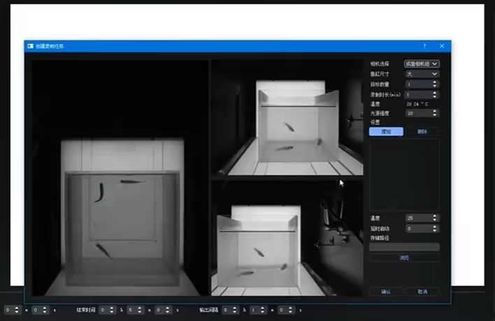
Why zebrafish imaging systems are essential for drug development timelines
In pharmaceutical R&D, a delayed decision is often more expensive than a slow instrument. A Zebrafish Imaging System helps teams turn a living-model observation into structured evidence while a compound series can still be adjusted. The value is not that one platform can predict every clinical outcome. A well-designed imaging workflow can instead make early phenotype, efficacy, and safety signals easier to capture, compare, and discuss.
For drug development teams, the critical question is not simply whether images look sharp. It is whether the Zebrafish Imaging System produces consistent readouts quickly enough to support a next experimental decision. When acquisition, orientation, analysis rules, and review are designed as one workflow, imaging can reduce avoidable waiting between dosing, observation, and interpretation.
The timeline problem is usually a data-handoff problem
Early discovery programs frequently lose time at the points between activities: compounds are prepared, embryos are dosed, images are captured, files are named, phenotypes are scored, and results are reconciled. A Zebrafish Imaging System does not eliminate biological uncertainty, but it can make those handoffs more repeatable. That matters when a project needs a defensible reason to advance, reformulate, repeat, or stop a candidate.
Zebrafish embryos are widely used in in vivo screening because they develop rapidly and are optically transparent. Those characteristics let researchers observe anatomy, fluorescent reporters, movement, vascular changes, or toxicity-associated phenotypes in a whole organism. Yet transparency alone does not create speed. If a lab relies on manual positioning, inconsistent exposure, and ad hoc image review, the analysis queue can become the real constraint on timelines.
A practical Zebrafish Imaging System therefore needs to be assessed as a decision-support workflow, not just as a camera. Its useful output is a comparable image set and a traceable quantitative or semi-quantitative conclusion.
How imaging shortens the path to a usable screening signal
1. It preserves biological context early
Cell-based assays can provide highly focused mechanistic information, while zebrafish add organism-level context. A Zebrafish Imaging System allows a team to inspect a phenotype in a living vertebrate model before committing every question to slower downstream studies. For example, brightfield and fluorescence readouts can be planned around morphology, reporter intensity, cell distribution, vessel patterning, cardiac motion, or behavior—depending on the assay design.
This does not replace mammalian studies or clinical evaluation. It can, however, make earlier drug development choices more informed by showing whether an intended effect is accompanied by a visible off-target or tolerability signal in the chosen zebrafish model.
2. It converts imaging from a viewing task into a measurement step
In published automated zebrafish workflows, image acquisition has been paired with quantitative analysis and orientation selection. That distinction is important. A Zebrafish Imaging System that only collects pictures can shift the bottleneck to manual scoring. A system paired with stable plate maps, controlled acquisition settings, and predefined analysis criteria can support more consistent comparisons across treatment groups.
For a laboratory, this means defining the endpoint before running a screen: what is measured, which image quality checks apply, how excluded embryos are recorded, and who reviews exceptions. This kind of discipline improves efficiency because investigators spend less time reinterpreting inconsistent image sets after the fact.
3. It makes repeat experiments easier to explain
Unexpected findings are common in discovery. The advantage of a configured Zebrafish Imaging System is not a guarantee that every repeat will agree; it is a clearer record of what was held constant and what changed. Standardized illumination, magnification, timing windows, sample orientation, and naming conventions make it easier to distinguish a biological difference from a workflow difference.
That traceability can protect timelines. Teams can decide whether a repeat is scientifically necessary instead of repeating an experiment because the original images or scoring logic cannot be reconstructed.
Where a Zebrafish Imaging System adds the most value
The strongest fit is usually an assay with a visible, relevant, and pre-specified endpoint. A Zebrafish Imaging System can be particularly useful for phenotype-based screening, developmental and organ toxicity studies, transgenic reporter assays, xenograft imaging, and cardiovascular or vascular observations. The appropriate configuration depends on the endpoint; a high frame rate may matter for cardiac motion, while fluorescence channels and analysis segmentation may matter more for labeled cells or reporters.
Recent imaging research also illustrates that advanced 3D approaches can expand what is observable. But a sophisticated modality is not automatically the efficient option. For routine drug development, a suitable setup matches the assay’s throughput, resolution, operator skill, and analysis capacity without introducing unnecessary handling or data-processing delay.
What to evaluate before you buy or configure a system
- Assay fit: Can the Zebrafish Imaging System capture the actual endpoint at the required time point and scale?
- Sample handling: How will plates, embryos, orientation, and failed wells be managed without hidden manual work?
- Image consistency: Are illumination, focus, exposure, and acquisition parameters stored and repeatable?
- Analysis workflow: Can the lab define scoring criteria, quality gates, and exception handling before the first screen?
- Data review: Can results be linked back to plate position, dose, image, and protocol version when a decision is questioned?
- Support for iteration: Does the supplier understand zebrafish applications well enough to help align the configuration with the assay rather than a generic specification sheet?
For many teams, this final point matters. Hunter Lab focuses on zebrafish instruments and applications and can support pre-purchase discussions about whether a proposed imaging approach suits a specific cardiovascular or phenotype assay. The right conversation should begin with the experimental question, not a promise of a fixed development-time reduction.
Build the workflow around decisions, not images
A Zebrafish Imaging System becomes valuable when it is embedded in a simple operating model: define the biological question, pre-register the image endpoint, standardize acquisition, apply a review rule, and connect the result to the next project decision. With that structure, a lab can use zebrafish imaging to identify which evidence needs confirmation and which candidates merit further investment.
That is why imaging is essential to drug development timelines: it helps make early in vivo evidence more accessible, comparable, and actionable. The gain is not a shortcut around validation. It is a more efficient route from observation to a decision the team can explain.








