
 EN
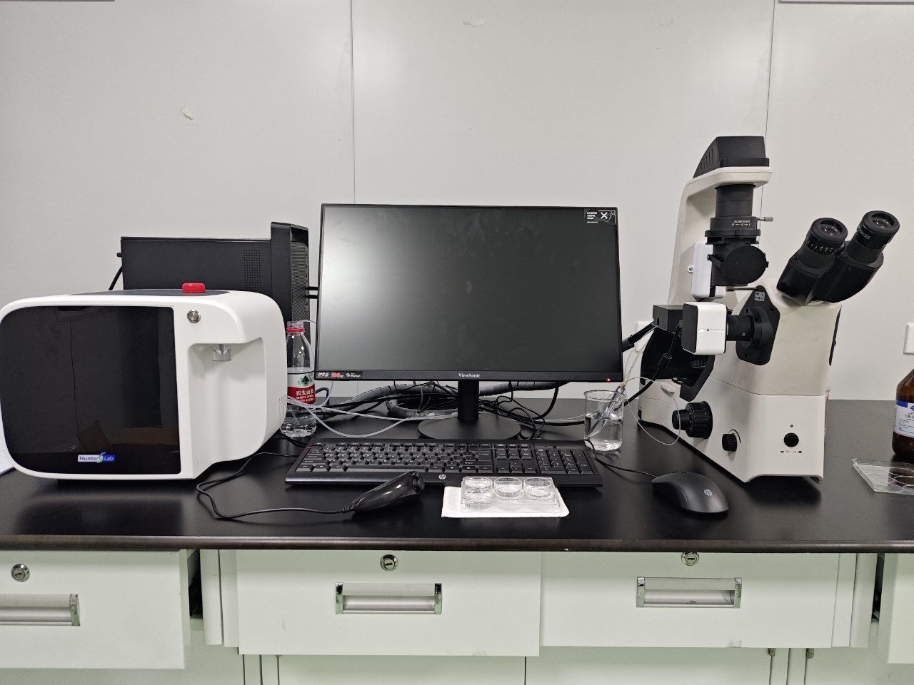
Why Is Zebrafish Fluorescence Imaging Essential for Drug Efficacy Studies?
In an efficacy study, seeing a zebrafish is not the same as measuring a response. The useful question is whether a treatment produces a reproducible biological change at the tissue, cell, pathway, or disease-model level. A Zebrafish Fluorescence Imaging System makes that change visible as a structured fluorescence readout, so a laboratory can move beyond a broad statement such as “the larvae looked better” and examine where, when, and how strongly the response occurred.
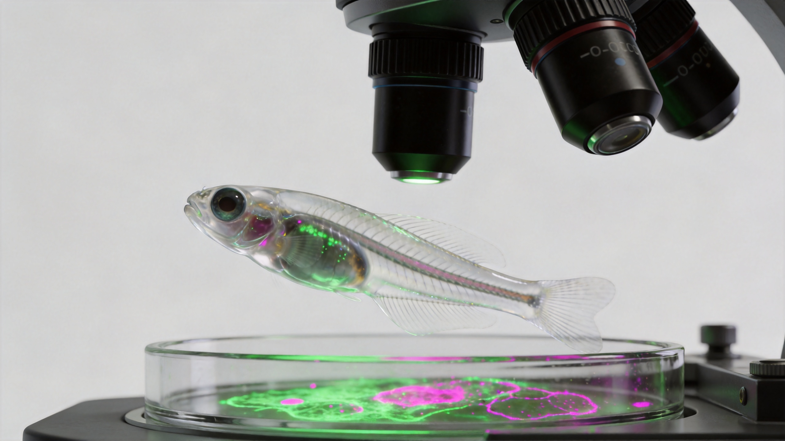
For teams screening candidate compounds, this distinction matters. Drug efficacy depends on evidence that a signal shifts in the intended direction while the experiment remains interpretable. Transparent embryos and larvae make zebrafish especially suitable for microscopy-based observation of labelled cells, organs, and processes in a living vertebrate. Fluorescent reporters, dyes, or labelled xenografts can turn a biological question into a measurable imaging endpoint without requiring the sample to be removed for every observation.
The problem: efficacy can be missed when the endpoint is too broad
Traditional observations such as survival, gross morphology, or simple behaviour can be valuable, but they may not reveal an early or localized pharmacological response. A compound may reduce inflammatory-cell recruitment, normalize vessel patterning, suppress a tumour-associated signal, or change an apoptosis marker before the whole animal shows an obvious difference. If the study relies only on a late, broad endpoint, a potentially useful response can be diluted by biological variability.
A Zebrafish Fluorescence Imaging System helps address that gap by capturing the signal in a defined region of interest. The same design can compare control and treated groups, quantify fluorescent area or intensity under a consistent acquisition method, and retain images for review. That does not make every fluorescence change proof of efficacy—signal changes can also reflect exposure, developmental delay, toxicity, imaging settings, or reporter biology—but it gives the study a more direct route to testing a mechanism-linked hypothesis.
Why fluorescence makes drug efficacy more interpretable
It connects the endpoint to a biological process
A well-chosen reporter is more informative than a decorative image. For example, fluorescence may label vasculature in an angiogenesis model, immune cells in an inflammation model, apoptotic cells in a safety or response assay, or a transplanted tumour population in an oncology workflow. The experimenter can then ask whether treatment changes the relevant readout in the expected anatomical location. Published zebrafish high-content workflows have described GFP-tagged cell counts, dual-fluorophore analysis, fluorescence-based angiogenesis measures, and apoptosis-oriented outputs, illustrating how distinct biological questions can be converted into imaging endpoints.
This specificity is central to drug efficacy. A reduction in overall fluorescence is not automatically a positive result. In a vascular assay, the meaningful comparison may be vessel-associated fluorescent area, branching pattern, or a defined region rather than whole-body brightness. In a cell-based reporter assay, the key signal may be cell number, distribution, or a ratio between channels. The endpoint needs to match the pharmacology question before a result can be interpreted responsibly.
It preserves spatial context
Bulk measurements can show that something changed, yet leave uncertainty about where the response occurred. Fluorescence imaging preserves spatial context: the signal can be associated with a target tissue, lesion, vessel bed, immune-cell population, or anatomical region. That context matters when two compounds produce a similar average intensity but affect different parts of the model. A Zebrafish Fluorescence Imaging System lets a team define and consistently apply regions of interest, making the evidence easier to review across plates and experiments.
Spatial context also helps identify misleading results. Diffuse background signal, irregular orientation, autofluorescence, or developmental abnormalities can be recognized in images in a way that a single aggregate number may hide. If the imaging room, strain, developmental stage, or exposure time changes, the calculation needs adjustment; a numerical output is only as comparable as the acquisition conditions behind it.
It supports time-aware pharmacology
Many efficacy questions are time dependent. A treatment may cause an early pathway response, a delayed tissue-level effect, or an effect that disappears after washout. Repeated or staged imaging allows the workflow to compare a baseline or vehicle condition with planned post-treatment windows. Rather than treating one image as a final verdict, researchers can build an evidence trail around onset, magnitude, and persistence of the fluorescence signal.
That is particularly useful when choosing dose and exposure windows. A strong signal shift at one time point may be less compelling if it coincides with general toxicity or developmental delay. Conversely, a modest but consistent change in the relevant fluorescent endpoint, accompanied by normal morphology and appropriate controls, can be more informative for prioritizing a candidate for follow-up pharmacology.
What a reliable fluorescence workflow needs
The value of a Zebrafish Fluorescence Imaging System comes from the workflow around it. A credible drug efficacy experiment begins with a biological question and a predefined endpoint, not with a search for attractive images. The following choices deserve attention:
- Reporter or label fit: select a fluorescent model or stain whose signal relates to the pathway, cell type, or phenotype being tested.
- Matched controls: include vehicle controls and, where appropriate, a known active or reference condition so that an observed shift has context.
- Consistent acquisition: lock exposure, magnification, channel selection, plate layout, orientation rules, and developmental timing as far as the assay allows.
- Predefined analysis: decide whether the readout is area, integrated intensity, object count, vessel-associated signal, or a multi-channel relationship before reviewing results.
- Quality review: document exclusions such as blurred wells, malformed larvae, saturation, or excessive background instead of silently mixing them into the analysis.
One practical lesson is that a bright signal is not necessarily a better assay. The metric that changes a protocol is often the one that reduces ambiguity: for one study it may be normalized fluorescent area; for another, a count of labelled cells within a defined region. When the literature and the bench disagree, check the strain, developmental stage, anaesthesia, mounting method, and imaging settings before attributing the discrepancy to the compound.
Fluorescence readouts can improve screening decisions
In an early screen, imaging can help separate compounds that merely alter appearance from candidates that affect the intended biology. A system designed for zebrafish work can support repeatable capture and analysis of fluorescence alongside brightfield context, which is helpful when teams need to inspect morphology while quantifying a molecular or cellular signal. Dedicated cardiovascular analysis workflows, for example, may combine visual assessment of heart motion with other assay-specific endpoints; the correct configuration depends on the model and the decision the data must support.
This does not replace confirmatory studies. Zebrafish data should be interpreted within the model’s limits and alongside exposure information, replicate structure, orthogonal endpoints, and later-stage testing. Still, when a Zebrafish Fluorescence Imaging System is paired with a suitable reporter and disciplined controls, it can make drug efficacy studies more selective, traceable, and biologically meaningful.
How to judge whether imaging is essential for your study
Fluorescence imaging is most valuable when the study needs an in vivo signal that is localized, repeatable, and closer to the mechanism than a broad phenotype alone. Ask three questions: Does the fluorescent marker represent the biology the compound is expected to change? Can the team standardize the image-acquisition and analysis rules? Will the resulting readout change a go/no-go, dose-selection, or follow-up decision?
If the answer is yes, fluorescence imaging is not simply an illustration layer. It is a method for converting a live zebrafish model into evidence that can be inspected, quantified, and challenged by the study team. That is why it has an important role in modern pharmacology: it helps connect a candidate compound to a visible, biologically relevant response while retaining the context needed to interpret that response carefully.
Frequently asked questions
Can fluorescence alone prove drug efficacy?
No. Fluorescence can provide a relevant quantitative endpoint, but the conclusion should be supported by appropriate controls, replicate design, exposure context, morphology review, and ideally orthogonal follow-up measurements.
What should be measured in a zebrafish fluorescence assay?
The metric should be predefined and tied to the biology: examples include fluorescent area, object count, intensity within a region of interest, signal distribution, or the relationship between two fluorescent channels.
Why use a zebrafish model for fluorescence-based screening?
Early zebrafish life stages are optically accessible, allowing researchers to observe labelled biological processes in a living vertebrate model. The appropriate assay choice still depends on the disease model, reporter, and pharmacological question.








