
 EN
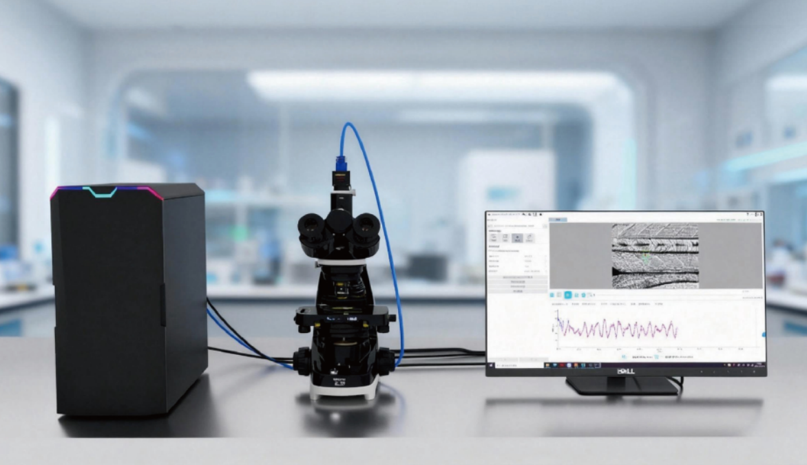
Why is a zebrafish imaging system essential for developmental biology research?
Developmental biology asks a deceptively difficult question: not only what changed, but when, where, and in what sequence did it change? A Zebrafish Imaging System turns that question into an observable workflow. Instead of relying solely on terminal snapshots, researchers can follow a living embryo through defined stages, record morphology and signal over time, and preserve the context needed to interpret a phenotype.
For teams working in developmental biology, the value is not simply a sharper picture. It is the ability to make imaging repeatable across embryos, operators, and experimental days. As researchers who work with live zebrafish assays know, a developmental observation is only as useful as the stage definition, acquisition conditions, and analysis path that travel with it.
Live observation makes developmental change measurable
Zebrafish are especially useful for live developmental work because early embryos and larvae are optically accessible. That accessibility lets researchers observe tissue boundaries, organ formation, cell movement, fluorescent reporters, and functional changes in the same organism. A Zebrafish Imaging System provides the practical bridge between that biological advantage and a dataset that can be compared later.
Time matters. Developmental stage is not interchangeable with a calendar label: temperature, strain, and husbandry can affect how a cohort progresses. For example, staging references commonly describe hatching over a range of roughly 48 to 72 hours post-fertilization under controlled conditions and place later landmark stages near 72 hours at 28.5 °C. Those values are useful context, not universal cutoffs. A robust imaging record should therefore capture the actual stage and conditions used for each embryo.
Without a consistent acquisition workflow, a difference in focus, orientation, exposure, or sampling interval can look like biology. With structured imaging, researchers can separate a genuine developmental trajectory from an avoidable technical variation.
Why standardization matters as much as resolution
High resolution is valuable only when it serves the biological question. In developmental biology, the more important requirement is often reproducibility: returning to the same anatomical region, using comparable contrast or fluorescence settings, and retaining metadata that explains how the image was acquired.
A common early protocol measures a visible endpoint once and treats it as the whole story. In practice, the metric that changes the protocol is often the rate and sequence of change. A delayed structure, for example, may look similar to a smaller structure in a single frame, yet a time series can reveal whether it is catching up, regressing, or showing no initiation during the observed period. That distinction changes both the hypothesis and the next assay.
- Stage-aware capture: organize each embryo by developmental stage, not only by clock time.
- Repeatable positioning: reduce variation in orientation and field of view so structures can be compared fairly.
- Stable acquisition settings: document illumination, objective, exposure, and fluorescence channels before comparing runs.
- Traceable analysis: keep image-processing choices tied to the original files and sample identifiers.
These practices matter when an effect is subtle. If two operators use different framing or thresholding rules, apparent phenotypic differences may reflect analysis choices rather than the embryo. A Zebrafish Imaging System supports a more disciplined chain from sample handling to quantification.
One system can connect morphology, function, and timing
Development rarely changes in a single dimension. An embryo may show altered body shape, a shifted fluorescent pattern, and a functional change that becomes visible only during a narrow time window. The advantage of integrated imaging is that those observations can be aligned rather than stored as separate, difficult-to-reconcile notes.
For cardiovascular development, a system that supports time-resolved acquisition can help a team examine cardiac motion alongside anatomy. For neural or cell-migration studies, controlled repeat imaging can reveal where a signal emerges and how it changes. The appropriate configuration depends heavily on the assay; there is no single setup that is right for every lab. Field of view, temporal resolution, fluorescence capability, sample throughput, and the downstream analysis burden should all match the biological endpoint.
Automation can be useful when a study must handle many embryos, but it should not remove biological judgement. Automated orientation or batch capture can improve consistency, while researchers still need to verify staging, exclude compromised specimens, and define meaningful endpoints before analysis begins.
How imaging strengthens phenotype interpretation
A developmental phenotype is more persuasive when it is anchored to a sequence rather than a single image. Consider an embryo with an apparently reduced organ size. Repeated imaging may show a late onset of growth, a transient developmental delay, or a structural defect that persists through the observed window. Those are different biological outcomes, even when a single endpoint image appears similar.
Imaging also helps teams decide when an assay needs refinement. A published protocol may recommend a particular interval, but in a local setup motion artefacts, embryo orientation, or reporter brightness can make that interval uninformative. Rather than forcing the dataset to fit the protocol, document the issue and adjust the acquisition plan with a clear rationale. This conditional approach is more useful than claiming that any one time point or instrument setting is universally optimal.
When results are reviewed later, the combination of original images, metadata, and repeatable measurements makes it easier to ask whether a finding is biologically coherent. That is essential for developmental biology, where cause and timing are closely linked.
A practical checklist for choosing a Zebrafish Imaging System
Before selecting a Zebrafish Imaging System, define the experimental decisions it must support. A system should be evaluated as part of a workflow, not as an isolated camera specification.
- Define the developmental question. Is the study tracking morphology, fluorescence, behavior, organ motion, or a combination?
- Set the imaging window. Specify the stages and sampling intervals that are meaningful for the phenotype.
- Match optics to the specimen. Balance resolution, depth, field of view, and gentle live imaging needs.
- Plan repeatability. Confirm how samples will be positioned, labeled, stored, and revisited across runs.
- Assess analysis early. Decide which measurements will be manual, automated, or reviewed by an experienced researcher.
- Test the whole workflow. Pilot a realistic cohort before scaling to a screen or longitudinal study.
Conclusion: imaging is experimental infrastructure, not an afterthought
A Zebrafish Imaging System is essential because it helps developmental biology move from compelling observations to interpretable evidence. It makes it practical to follow an embryo over time, standardize acquisition, preserve the conditions behind each image, and connect visible change to a defined developmental question.
The most effective workflow is not necessarily the one with the most complex hardware. It is the one that reliably captures the right information at the right developmental stage and gives the research team confidence that differences between embryos reflect biology rather than inconsistent imaging. For studies built around timing, morphology, and living processes, that consistency is foundational.








