
 EN
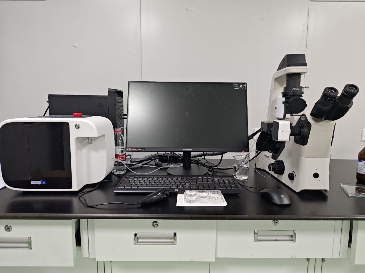
What resolution can a modern zebrafish imaging system achieve?
A modern Zebrafish Imaging System can produce anything from whole-larva documentation at micrometer-scale sampling to submicron optical detail in a tightly defined field of view. That range is wide because “resolution” is not one specification. It reflects the objective, wavelength, numerical aperture, camera pixel size, specimen depth, contrast, motion, and the way images are analyzed. For a lab choosing a system, the useful question is not “What is the highest resolution?” but “What resolution is reliable for the biological feature and throughput we need?”
From a shared-facility perspective, the strongest imaging workflows begin by matching the required detail to the assay. A whole-embryo morphology screen, a beating-heart measurement, and a fluorescence readout from a small tissue region each place different demands on pixel sampling, magnification, speed, and image quality.
Start by separating optical resolution from image resolution
The camera’s pixel count is easy to compare, but it is not the same as optical resolution. Optical resolution is the smallest separation between two nearby features that the microscope can distinguish. A commonly used Rayleigh estimate is approximately 0.61 times the illumination wavelength divided by the objective’s numerical aperture (NA). In practical terms, shorter wavelengths and a higher-NA objective can reveal finer detail, provided the sample, illumination, and detector support it.
A Zebrafish Imaging System may display a very large image while still failing to resolve a smaller biological feature. Enlarging an image adds pixels to the display; it does not add information beyond the optical limit. For that reason, a useful specification review should consider objective NA and effective sample-plane pixel size alongside sensor resolution.
What resolution is realistic for common zebrafish tasks?
No single number describes every Zebrafish Imaging System. The field of view creates the first trade-off. Whole larvae need a broad field and convenient working distance, which often favors lower magnification and coarser sampling. A localized fluorescence signal or fine anatomical boundary may justify a smaller field and a higher-NA objective.
- Whole-larva brightfield or morphology screening: The goal is typically robust visualization of body shape, gross edema, pigmentation, swim-bladder status, or obvious malformations. Consistent orientation and contrast can matter more than the smallest possible pixel size.
- Organ-level live imaging: Cardiac, vascular, or regional fluorescence work often needs a balance of spatial detail and frame rate. A higher magnification can clarify boundaries, but motion blur, lower field coverage, and slower acquisition may offset that gain.
- Fine fluorescence or cellular-scale work: Here, high NA, appropriate illumination, stable mounting, and carefully matched pixel sampling become more important. Depth, scattering, and photobleaching may still limit usable quality in a living specimen.
So the achievable resolution should be stated as an assay-dependent capability, not as a universal promise. For example, a system optimized for rapid imaging of many larvae may deliver very useful organ-level information without being the right configuration for resolving the smallest subcellular structure.
Why pixel size and magnification must be matched
Sampling determines whether the camera records the detail that the optics can provide. A practical microscopy principle is to sample the optical resolution with multiple camera pixels rather than one large pixel. One technical example places a roughly 250 nm optical resolution limit alongside a final image pixel size of about 80–100 nm. The exact value changes with the objective, camera, coupler, wavelength, and imaging mode, but the lesson is stable: sensor resolution alone does not determine image quality.
Have you calculated the effective sample-plane pixel size for the objective and camera you plan to use? The calculation is straightforward: divide the detector pixel dimension by the total optical magnification. Then compare that result with the detail you need to measure. If the pixels are too large, fine information is undersampled. If they are much smaller than the optical limit, files become heavier and acquisition may become slower without producing proportionally more useful detail.
Four limits that can reduce usable resolution
1. Numerical aperture and wavelength
Two objectives with the same stated magnification can produce different resolving power because their NA differs. Higher NA generally improves lateral optical resolution and light collection, although working distance and depth tolerance may change. The objective should therefore be evaluated as an optical component, not merely by its magnification label.
2. Specimen depth and contrast
Live zebrafish are optically favorable in many early developmental applications, yet depth, pigmentation, scattering, and out-of-focus fluorescence still affect image quality. A sharp surface image does not guarantee the same resolution deeper in the specimen. Matching the modality to the depth and signal type is usually more productive than simply choosing a camera with more pixels.
3. Motion and acquisition speed
For moving organs or behavioral assays, exposure time and frame rate can be the limiting factors. A technically finer image that blurs during cardiac motion is less informative than a slightly coarser image acquired consistently. That is the speed-versus-reproducibility trade-off many labs face when configuring a Zebrafish Imaging System.
4. Mounting, orientation, and focus consistency
The detail the camera catches can be lost before analysis if larvae are tilted, unevenly positioned, or focused at different depths. Automated positioning can improve repeatability for high-throughput imaging, but the workflow still needs a verification step for focus, orientation, and exposure. Good data are rarely created by resolution alone.
A decision framework for specifying a Zebrafish Imaging System
- Name the biological feature. Define its approximate size, location, contrast mechanism, and whether it moves.
- Set the minimum usable resolution. Decide what separation or boundary must be measured, not simply what looks sharp on screen.
- Choose the field of view first. Whole-larva coverage and detailed regional imaging require different optical trade-offs.
- Match pixel sampling to the objective. Confirm effective pixel size after the complete optical path, including adapters or binning.
- Test image quality under real assay conditions. Use the intended developmental stage, mounting method, illumination, and exposure settings rather than a static demonstration sample.
- Validate repeatability. Check whether different operators and imaging sessions produce comparable focus, orientation, and measurements.
The practical answer
A modern Zebrafish Imaging System can achieve impressive resolution, but its meaningful limit is defined by the complete workflow. For broad phenotyping, the right answer may be stable whole-organism imaging with adequate pixel sampling. For fine fluorescence or anatomy, it may require higher NA, controlled illumination, precise focus, and a narrower field. In many labs, the most suitable system is not the one that advertises the largest image; it is the one that produces repeatable, assay-appropriate quality at the needed throughput.
When evaluating options, ask vendors and internal users to demonstrate the exact combination of resolution, pixel size, magnification, field of view, and acquisition speed on a representative zebrafish assay. That evidence will tell you much more than a single headline specification.








