
 EN
A Zebrafish Cardiovascular Analysis System usually combines optics, controlled illumination, image capture, and quantification software rather than relying on one imaging method. The useful modality depends on the question: is the study measuring beat timing, chamber motion, red-cell movement, vessel morphology, fluorescent reporter expression, or three-dimensional structure? For early-stage larvae, optical clarity makes several approaches practical. For older fish, pigmented tissues, or deeper anatomy, the trade-offs change.
From a cardiovascular research perspective, the sound starting point is not “which camera is most advanced?” It is “what biological event must be visible, how quickly does it change, and what evidence will make the measurement defensible?” A well-configured Zebrafish Cardiovascular Analysis System links those choices to a repeatable protocol, including specimen orientation, temperature control, illumination, exposure time, and interpretation rules.
Brightfield and transmitted-light video microscopy
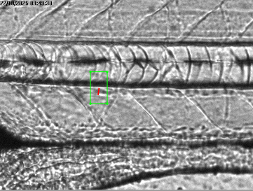
Brightfield or transmitted-light imaging is the everyday foundation of many workflows. With a stereomicroscope or microscope objective, a camera records contrast created by the embryo, heart wall, and moving blood cells. In this label-free configuration, the approach can be useful when the study needs routine morphology, heart-rate estimation, chamber contraction, gross circulation, or a rapid screen across many larvae.
Video microscopy matters because the zebrafish heart and circulation are dynamic. A still image may show anatomy, but it cannot establish the timing of systole and diastole or show whether moving cells are continuous, intermittent, or diverted. High-speed video microscopy has been used in zebrafish embryo studies of cardiac function and blood-flow hemodynamics. In practice, the necessary frame rate depends on the endpoint, developmental stage, magnification, and measurement method; it should be demonstrated during method validation rather than borrowed from an unrelated protocol.
For many laboratories, a Zebrafish Cardiovascular Analysis System built around transmitted light is a practical baseline because it avoids the need for fluorescent labeling. Its limits are equally important: weak contrast, motion blur, variable specimen position, and overlapping structures can affect segmentation. The workflow therefore benefits from stable mounting, consistent focus, controlled light intensity, and a predefined rule for excluding unusable recordings.
Fluorescence imaging for structures and reporter-defined signals
Fluorescence imaging adds specificity when a cardiovascular question concerns a labeled cell population, vessel network, or molecular reporter. Transgenic fish with tissue-specific fluorescent signals can make endothelial patterns, myocardial structures, or circulating cells easier to distinguish from surrounding tissue. In this workflow, fluorescence is particularly helpful when transmitted light shows motion but cannot clearly separate the feature being measured.
Widefield fluorescence microscopy can support two-dimensional observation of labeled structures. Confocal imaging can be considered when optical sectioning and finer spatial discrimination are important. Neither choice is automatically required: additional optical complexity can reduce throughput and may increase exposure-management concerns. Researchers should decide whether the added contrast answers a real question that a simpler image cannot.
Light-sheet fluorescence microscopy is another option for volumetric or developmental work. It can be valuable where a study needs repeated views of a fluorescent vascular network or a beating cardiac structure across depth. Yet a light-sheet workflow also needs careful attention to specimen mounting, acquisition timing, data volume, and motion correction. The image is only part of the assay; processing choices determine whether measurements remain comparable from fish to fish.
High-speed imaging and quantitative motion measurement
High-speed capture is often described as a separate modality, although it is more accurately a capability that can be paired with brightfield or fluorescence. In this setup, temporal resolution enables frame-by-frame analysis of cardiac cycles and moving blood cells. This supports endpoints such as beat interval, chamber-area change, wall-motion patterns, and relative flow behavior when the recording and algorithm have been validated for the experiment.
The key question is whether every part of the measurement chain resolves the event of interest. Short exposure can reduce blur, but it may require more illumination. Higher magnification can reveal a region of interest, but it reduces the field of view and may make tracking more sensitive to movement. A useful protocol documents camera settings, orientation, region-of-interest definitions, and the way failed tracks are handled. That detail is often more informative than simply labeling a system “high resolution.”
Optical coherence tomography and other depth-resolved options
Optical coherence tomography (OCT) is a non-invasive, depth-resolved optical technique that may extend what a microscopy workflow can reveal beyond surface-visible features. It can be relevant when researchers need cross-sectional context or when depth makes conventional microscopy less informative. Reviews of zebrafish cardiovascular applications describe OCT as a promising route for examining cardiac and vascular structures, including early developmental studies.
OCT does not replace every microscopy workflow. It has different contrast mechanisms, sampling geometry, acquisition requirements, and interpretation considerations. Its value rises when depth-resolved anatomy changes the decision a researcher can make; otherwise, standard video microscopy may be more efficient. This is a useful correction to a common assumption: more complex imaging is not inherently more suitable for a cardiovascular endpoint.
How to choose modalities within one workflow
Many studies benefit from a staged rather than single-modality design. A coordinated workflow may use brightfield video for primary screening, fluorescence to clarify a labeled vascular or cardiac feature in selected samples, and OCT or another depth-resolved method for a focused follow-up question. The modalities should be linked by a shared specimen-identification scheme and by endpoints defined before data collection.
| Question | Useful imaging approach | Practical consideration |
|---|---|---|
| Is the heart beating regularly? | Brightfield or transmitted-light video microscopy | Validate timing and exposure settings for the developmental stage. |
| How does visible blood-cell motion behave? | High-speed label-free video microscopy | Standardize orientation and tracking regions. |
| Where are labeled vessels or cardiac cells? | Widefield fluorescence, confocal, or light-sheet fluorescence | Confirm reporter suitability and manage photobleaching. |
| Is depth-resolved structure needed? | OCT or another depth-resolved optical method | Use it when cross-sectional information changes interpretation. |
What makes the images analytically useful?
Imaging becomes quantitative interpretation only when acquisition and interpretation are controlled together. Before deployment, define the primary endpoint, select a modality that can actually resolve it, and test reproducibility across operators or sessions. Record environmental conditions that may affect cardiovascular behavior, such as developmental timing and temperature. If automated segmentation is used, retain a review process for low-confidence outputs rather than assuming every detected contour is biologically meaningful.
For teams comparing configurations, Hunter Lab’s cardiovascular application expertise can help frame the conversation around assay requirements rather than a generic equipment checklist. The appropriate configuration is the one whose imaging, motion capture, and measurement workflow fit the study’s intended endpoint and can be transparently validated.
Conclusion
The principal modalities are brightfield or transmitted-light video microscopy, fluorescence imaging, high-speed acquisition paired with either contrast method, and depth-resolved methods such as OCT. Each modality contributes a different kind of evidence. Select the simplest modality that resolves the relevant event, then document the acquisition and processing choices that make the resulting cardiovascular measurements interpretable.








