
 EN
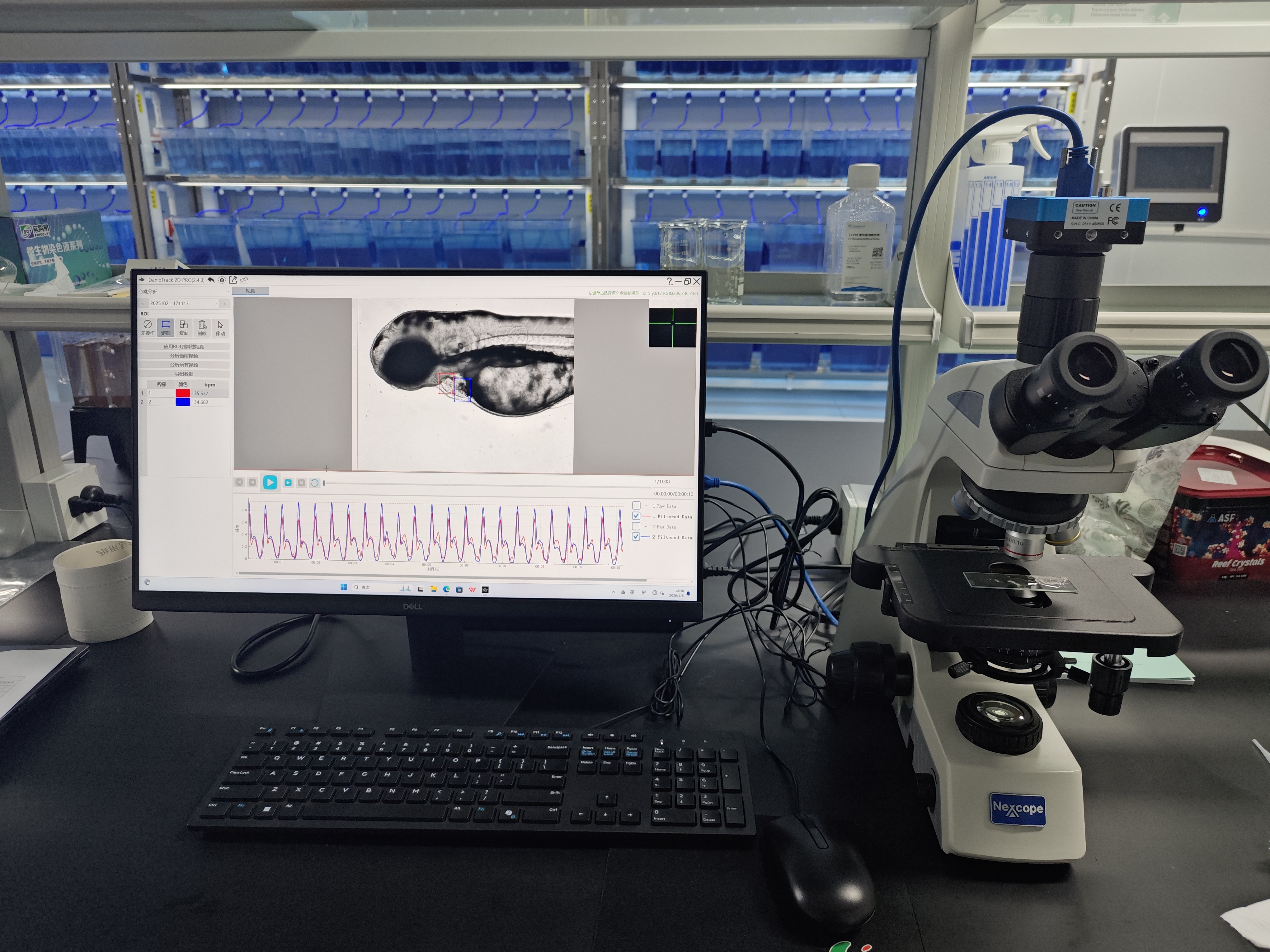
A Zebrafish Imaging System can bring several complementary views of the same living model into one workflow. Rather than treating brightfield, fluorescence, confocal, and volumetric acquisition as interchangeable settings, a multimodal setup uses each mode for the question it answers well: morphology, labeled biology, three-dimensional structure, or time-resolved function. This matters because young zebrafish are optically transparent enough for in vivo observation, yet a single image type rarely captures every relevant change.
For a research team planning a Zebrafish Imaging System, the useful question is not simply “which modality has the highest specification?” It is whether the system can produce linked observations at the developmental stage, field of view, and acquisition speed that an assay requires. A change that looks subtle in transmitted light may become interpretable when it is compared with fluorescence distribution or a three-dimensional optical section.
What multi-modal imaging means in zebrafish research
Multi-modal imaging combines two or more imaging methods so that they describe different features of the same specimen or experimental series. In zebrafish work, that often means pairing low-magnification brightfield imaging with fluorescence acquisition, then adding confocal or light-sheet imaging when depth information is important. The value comes from correspondence: images should be collected under an aligned sample identity, orientation, timing plan, and analysis workflow.
A practical Zebrafish Imaging System may support the following capability layers:
- Transmitted-light or brightfield imaging for gross anatomy, development, pigmentation, edema, body axis, and other visible phenotypes.
- Fluorescence imaging for reporter lines, labeled cells, tissues, vascular patterns, or fluorescent assay signals.
- Optical sectioning, often through confocal methods, when out-of-focus signal would obscure a structure of interest.
- Volumetric imaging, including light-sheet approaches, when a researcher needs a three-dimensional view of a larger region or a dynamic process across depth.
- Time-lapse acquisition and analysis for developmental, cardiac, behavioral, or reporter-signal changes that cannot be inferred from a single endpoint.
The modes do not all need to be used in every experiment. A screening assay may begin with rapid imaging of morphology and add fluorescence only for samples that meet a predefined review criterion. A developmental study may use brightfield to document stage and orientation, then use a deeper modality to inspect a labeled tissue. That staged design is often more useful than collecting maximum-detail data from every larva.
Brightfield provides a fast structural baseline
Brightfield or transmitted-light imaging gives the broadest visual context. It is commonly used to verify that a larva is positioned consistently, to document gross morphology, and to identify sample-quality issues before a more demanding acquisition begins. In a multi-modal workflow, this baseline image can also help analysts interpret a fluorescence pattern in its anatomical context.
For example, a fluorescence signal near the trunk is easier to evaluate when a corresponding brightfield image shows body orientation, curvature, and focal plane. Brightfield does not identify every biological mechanism, but it can make an observation auditable by showing what the specimen looked like at the time of acquisition. That is a key capability for teams building reproducible records across repeated imaging sessions.
Fluorescence adds cell, tissue, and functional contrast
Fluorescence turns a Zebrafish Imaging System into a tool for targeted biological questions. Transgenic reporters, fluorescent dyes, and labeled structures can reveal where a signal is present and how it changes over time. Depending on the assay, fluorescence may be used to examine vascular architecture, tissue-specific expression, inflammatory-cell distribution, calcium-related activity, or changes in an assay-specific marker.
The practical limitation is that fluorescence images must be acquired and interpreted with attention to exposure, channel separation, photobleaching, and sample movement. If the goal is comparison across plates or days, the acquisition plan should define the same channel settings, developmental window, mounting approach, and quality controls. Multi-modal work is strongest when the fluorescence result is paired with a structural image rather than assessed in isolation.
Confocal imaging clarifies structure through optical sectioning
Confocal imaging is useful when a widefield fluorescence image contains overlapping signal from different depths. By collecting optical sections, it can help researchers inspect finer relationships within a region of interest and build a z-stack for later review. In zebrafish studies, that can be relevant for labeled vessels, localized reporter expression, cardiac structures, or cell distributions where depth matters.
This added detail has a trade-off: optical sectioning can require more acquisition time and may be less suitable for every high-throughput experiment. The right decision depends on whether the study needs a detailed three-dimensional answer or a rapid, repeatable screening readout. If throughput is the binding constraint, a faster two-dimensional screen may be appropriate; if depth-dependent structure is the question, confocal acquisition becomes more valuable.
Light-sheet and volumetric imaging broaden the field of view
Light-sheet fluorescence approaches illuminate a thin plane rather than exposing the full sample volume at once. In published zebrafish work, light-sheet methods have been used for efficient live imaging of large regions, including the heads of transgenic embryos, and complementary light-sheet and tomography workflows have documented whole embryos over developmental time. These examples illustrate why volumetric methods are relevant when the biological event extends across depth or a broad anatomical region.
For a Zebrafish Imaging System, volumetric capability is not a reason to abandon simpler modes. It is a way to escalate when a two-dimensional view cannot answer the question. The imaging plan should define the volume, interval, and signal needed before acquisition starts. More planes do not automatically yield more useful data if the sample orientation, motion control, or analysis capacity is not ready for the resulting dataset.
Time-lapse imaging connects structure to change
One important advantage of live zebrafish imaging is the ability to observe change rather than comparing only separate endpoints. Time-lapse sequences can track developmental progression, reporter dynamics, circulation-related movement, or recovery after a defined experimental event. The design challenge is to balance temporal sampling with light exposure, data volume, and the stability of the sample environment.
For long sessions, treat temperature, anesthesia or immobilization approach, light schedule, and mounting as controlled variables. Published baseline values are not universal constants: they depend on the biological stage and local conditions. A Zebrafish Imaging System supports meaningful time-series work only when the associated protocol documents those conditions alongside the images.
How modalities work together in one workflow
A clear multi-modal workflow usually follows a deliberate sequence:
- Define the biological decision. State whether the study is looking for morphology, a fluorescent marker, three-dimensional organization, dynamic change, or a combination.
- Set the structural baseline. Capture brightfield images for orientation, specimen condition, and broad phenotype review.
- Add targeted contrast. Use fluorescence channels only where a reporter or label answers a defined question.
- Escalate to depth-resolved acquisition when needed. Use confocal or volumetric methods when superimposed signal or anatomy across depth changes the interpretation.
- Plan analysis before collection. Name files consistently, retain acquisition metadata, and decide how structural and fluorescent observations will be registered or compared.
This sequence supports versatility without turning every project into an oversized imaging exercise. It also helps a team identify which part of the workflow is limiting: sample preparation, optics, acquisition speed, or image analysis.
Questions to ask when evaluating imaging capabilities
When assessing a Zebrafish Imaging System, start with your assay rather than a generic modality list. Ask whether the intended workflow needs whole-larva context, tissue-specific contrast, optical sections, rapid repeated acquisition, automated positioning, or standardized analysis. Then verify how the platform handles channel changes, z-stacks, multiwell formats, metadata, and repeatability between operators.
Hunter Lab focuses its product line on zebrafish instruments and applications, but the appropriate configuration still depends on the study design. The most useful system is the one that links the required imaging modes to a controlled sample workflow and a defensible analysis plan. For a lab that needs broad screening, fast structural and fluorescence review may be the starting point. For a lab studying depth-dependent signals or live developmental processes, confocal or volumetric capabilities may deserve greater weight.
Conclusion
The multi-modal value of a Zebrafish Imaging System lies in combining complementary evidence: brightfield context, fluorescence specificity, depth-resolved structure, and time-aware observation. Select the modes that answer your actual biological question, keep sample handling and acquisition conditions consistent, and make sure the analysis workflow can connect the resulting datasets. That approach turns multi-modal imaging from a feature list into a reliable research method.








