
 EN
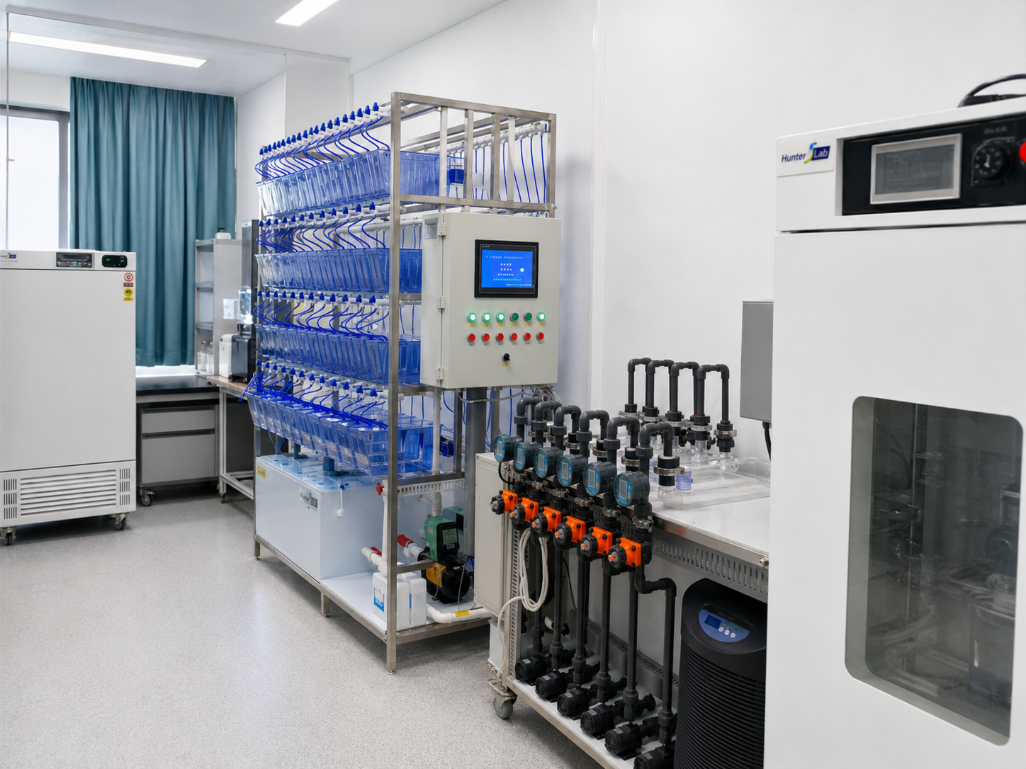
Cardiovascular assays in zebrafish are valuable because early life stages are optically accessible and cardiac activity can be observed in a living organism. Yet a useful assay is not just a matter of collecting videos. Laboratories also need a repeatable way to acquire images, define measurements, retain the raw record, and compare one plate or study with another. A Zebrafish Cardiovascular Analysis System brings those steps into a more structured workflow. When the workflow is validated for the strain, developmental stage, temperature, and study question, automation can make cardiac phenotyping easier to scale and easier to review.
For researchers planning drug-safety screens, developmental studies, or disease-model experiments, the main benefit is not that a system replaces scientific judgment. It is that a Zebrafish Cardiovascular Analysis System can apply the same acquisition and analysis logic across many samples. That consistency matters when small changes in handling, focus, timing, or manual scoring could obscure a biological signal.
Why manual cardiovascular assessment becomes a bottleneck
Manual review can be appropriate for a small pilot experiment or an unusual phenotype. It becomes harder to manage when a study has many wells, repeated time points, or multiple analysts. One operator may select a different cardiac cycle, draw a region of interest differently, or record an observation in a separate spreadsheet. Those choices can be reasonable, but they make comparison harder if they are undocumented.
As a researcher working with zebrafish cardiovascular studies, I have seen how a protocol can appear stable until data from two imaging sessions must be pooled. We started out measuring heart rate alone, thinking it was the most direct indicator, but once we looked at rhythm, wall motion, and flow-related observations alongside it, the interpretation became more useful. A Zebrafish Cardiovascular Analysis System helps organize that broader view by connecting acquisition to parameter extraction and review.
1. More consistent image acquisition and measurement rules
Automation can standardize repeatable actions such as plate handling, image-capture timing, exposure settings, focus routines, naming conventions, and calculation templates. In a validated Zebrafish Cardiovascular Analysis System, those settings are defined before the run rather than reconstructed later from notes. This supports consistency across samples and makes deviations easier to identify.
That does not mean every experiment should use identical settings. A larval developmental study and an adult physiology study may require different imaging conditions. The advantage is that a system can retain the approved method for each assay rather than relying on memory. If room temperature, anesthesia exposure, or embryo orientation changes, the laboratory can document the condition and interpret the result in context.
2. Greater throughput without losing the audit trail
Cardiac imaging generates a substantial amount of information: video frames, acquisition metadata, parameter outputs, quality flags, and reviewer decisions. A Zebrafish Cardiovascular Analysis System can support throughput by moving repetitive capture and first-pass analysis into a defined sequence. Researchers can then devote more time to reviewing exceptions, confirming biologically meaningful changes, and refining experimental design.
Published automated imaging workflows have shown that heartbeat-related information can be extracted from zebrafish imaging data at scale. The relevant question for a laboratory is whether its own workflow maintains usable image quality and clear quality-control rules. A Zebrafish Cardiovascular Analysis System should make it possible to flag recordings with motion artifacts, poor orientation, or insufficient contrast rather than treating every output as equally reliable.
3. Simultaneous, multi-parameter cardiovascular context
A visible phenotype is often more informative when it is considered with other measurements. Depending on the validated method, a Zebrafish Cardiovascular Analysis System may organize observations such as heart rate, beat-to-beat regularity, ventricular motion, fractional shortening, vessel diameter, or blood-flow behavior. The value is the relationship among the measurements, not a single isolated number.
For example, a heart-rate shift may be associated with developmental delay, handling conditions, rhythm disturbance, or a compound response. A workflow that preserves the source recording and the analysis settings lets the team revisit the evidence. This is especially useful in borderline findings, where automated output should prompt informed review rather than an automatic conclusion.
4. Better study-to-study comparability
Many cardiovascular programs span weeks or months. In that setting, efficiency includes the ability to retrieve prior settings, compare batches, and understand why a result changed. A Zebrafish Cardiovascular Analysis System can associate data with plate maps, dates, assay versions, and review outcomes. That structure gives a team a clearer basis for deciding whether a difference is likely biological or whether it needs a method check.
Consistent reporting also supports collaboration. A core facility can provide investigators with a defined output package rather than a mix of videos, handwritten observations, and separate spreadsheets. For university laboratories and research groups working across projects, this can make handoffs more straightforward while keeping the scientific questions visible.
5. A practical foundation for screening decisions
Automated cardiovascular analysis is particularly useful when researchers need to prioritize samples for further investigation. A Zebrafish Cardiovascular Analysis System can help sort large sets of recordings into review categories using pre-agreed criteria. It does not establish clinical safety, and it should not be treated as a substitute for follow-on models or domain expertise. Instead, it supplies a more disciplined evidence layer for deciding what merits the next experiment.
This distinction is important in preclinical work. Automation improves efficiency when the method is matched to the decision: exploratory phenotype discovery may need broad capture and flexible review, while a repeat screen may need tighter acceptance criteria. The right configuration depends heavily on the assay endpoint and the laboratory’s validation plan.
What to evaluate before adopting an automated workflow
- Fit to the endpoint: Define whether the study needs rate, rhythm, contractility, flow, morphology, or a combination.
- Image quality controls: Establish rules for focus, orientation, motion artifacts, and excluded samples before analysis begins.
- Method traceability: Confirm that acquisition settings, analysis settings, and reviewer decisions can be retained with the result.
- Reproducibility checks: Compare repeat plates and operators under controlled conditions before using outputs for major decisions.
- Human review: Plan how experts will inspect outliers and ambiguous recordings rather than relying on an output alone.
Conclusion
The core advantages of a Zebrafish Cardiovascular Analysis System are more consistent acquisition, practical throughput, richer multi-parameter context, and a clearer data trail for review. Automation is most valuable when it is paired with a well-defined protocol and thoughtful quality control. For teams studying zebrafish cardiac phenotypes, that combination can turn a collection of imaging observations into a more structured, repeatable research workflow.








