
 EN
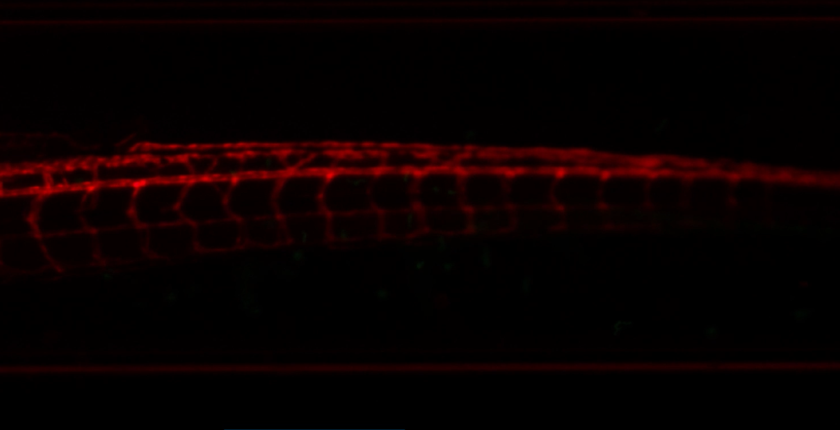
The zebrafish fluorescence imaging system supports both fixed and live sample imaging.
A Zebrafish Fluorescence Imaging System can support two complementary research routes: a fixed sample workflow for endpoint detail and a live sample workflow for observing change over time. That adaptability matters because a question about marker location, tissue architecture, or phenotype confirmation is not the same as a question about cellular movement, circulation, or treatment response. The most useful choice is therefore not “fixed or live” in the abstract. It is matching the acquisition plan to the biological event that must be measured.
For researchers setting up a new assay, the practical value of a Zebrafish Fluorescence Imaging System is its ability to keep both routes available within one acquisition strategy. Zebrafish embryos and early larvae are optically accessible, which makes fluorescence microscopy a practical way to examine labeled cells and tissues. Still, accessibility does not remove the need to define the endpoint before mounting a sample.
Why fixed and live observation answer different questions
fixed-sample microscopy is an endpoint method. A specimen is preserved before staining or observation, so the workflow can prioritize marker specificity, anatomical orientation, and consistent comparison across a batch. Whole-mount immunofluorescence is widely used for microscopy-based phenotypic analysis in zebrafish. With a Zebrafish Fluorescence Imaging System, fixed preparations are especially useful when a team needs to document where a signal appears after treatment, compare tissues across groups, or retain a stable specimen for repeated acquisition settings.
living specimen microscopy, in contrast, is built around time. It can reveal a fluorescent pattern as development, migration, circulation, or another biological process unfolds. A Zebrafish Fluorescence Imaging System used for a living specimen must balance signal collection against the realities of a living specimen: motion, mounting, temperature stability, and the duration of acquisition. That does not make live observation less reliable; it means the acquisition plan needs to be designed around the event being observed.
What fixed-sample microscopy makes easier
A preserved specimen workflow generally gives the operator more control over preparation and acquisition timing. Because the specimen is no longer changing, a Zebrafish Fluorescence Imaging System can be used to collect comparable fields across groups without trying to synchronize a transient event. This is helpful for endpoint phenotyping, localization studies, and fluorescence assays that depend on post-fixation labeling.
- Stable anatomy: a preserved specimen can be positioned carefully for repeatable views of a region of interest.
- Marker-focused readout: staining and microscopy can be optimized around the target signal rather than around the survival window of a live animal.
- Batch comparison: consistent illumination, exposure, and orientation make it easier to compare groups when the protocol is standardized.
The trade-off is straightforward: a preserved specimen records a final state, not a process. If the research question depends on when a signal appears or how it changes, a Zebrafish Fluorescence Imaging System should be planned for live acquisition as well.
What living specimen observing adds
living specimen work turns fluorescence from a static marker into a temporal readout. A Zebrafish Fluorescence Imaging System can support time-lapse or repeated observations when the study asks how a signal evolves in the same animal. This is valuable for developmental observations and for experiments in which the timing of a response matters as much as its final intensity.
Long-term live observation is demanding, however. Movement can blur a signal or complicate tracking, and prolonged acquisition can change the practical limits of a session. In practice, the first instinct may be to collect the longest possible sequence. In reality, a shorter sequence with a clearly defined biological window often produces a more interpretable result. The right schedule depends heavily on developmental stage, fluorescent label, mounting method, and the question being tested.
- Time-resolved observation: a living specimen can show onset, progression, or recovery rather than only an endpoint.
- Within-sample comparison: repeated observations may reduce ambiguity when the same specimen is followed over time.
- Protocol sensitivity: mounting, motion control, focus stability, and acquisition intervals should be piloted before scaling up.
For this reason, a Zebrafish Fluorescence Imaging System is most versatile when the operator can move between fixed and living specimen workflows without treating one as a substitute for the other.
A practical decision framework
Start with the endpoint. If the study needs a high-confidence map of a fluorescent marker after a defined treatment or developmental stage, a preserved specimen approach is often the cleaner first choice. If the study needs to know how that marker changes, moves, or responds over time, a living specimen plan becomes more appropriate. A Zebrafish Fluorescence Imaging System supports this decision by providing the microscopy foundation; the protocol determines whether the output is meaningful.
| Research need | More suitable workflow | Key planning question |
|---|---|---|
| Endpoint localization or morphology | preserved specimen | Which marker and orientation most directly answer the question? |
| Dynamic developmental or treatment response | living specimen | What time window and motion-control plan preserve interpretability? |
| Method confirmation before a dynamic study | preserved specimen followed by living specimen | Can the endpoint signal validate the target before time-lapse work? |
How to plan a versatile fluorescence workflow
A Zebrafish Fluorescence Imaging System is most useful when the acquisition plan is written before specimens are prepared. Define the fluorescent target, developmental stage, field of view, expected timing, and the comparison that will be made. Then decide whether the experiment needs a preserved specimen, a living specimen, or both. This small planning step prevents a common problem: collecting attractive images that do not directly answer the experiment’s primary question.
It also helps to run a modest pilot. Use it to check focus, background, sample orientation, and whether the acquisition interval fits the biological process. A pilot is not merely a technical rehearsal. It is where the workflow’s adaptability becomes visible: the same Zebrafish Fluorescence Imaging System can support a preserved specimen confirmation step and a carefully controlled living specimen observation, provided each protocol is designed for its own purpose.
Conclusion
The strength of a Zebrafish Fluorescence Imaging System is not that it makes fixed and living specimen microscopy identical. Its strength is that it supports both, allowing researchers to choose endpoint clarity, dynamic observation, or a combined workflow based on the biological question. Fixed samples provide stable, marker-focused evidence. Live samples add timing and behavior. When that distinction guides the protocol, fluorescence microscopy becomes a more versatile and defensible part of zebrafish research.








