
 EN
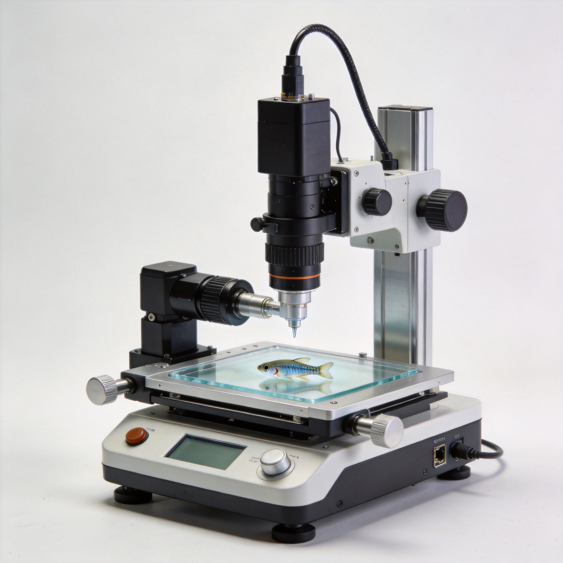
Modern zebrafish imaging systems feature automated focus and stage control for efficiency.
In a busy core facility, time is seldom lost in one dramatic failure. More often, it slips away in the pauses between wells: refocusing after a field change, returning to a coordinate, checking whether a larva drifted, or deciding whether a soft image should be repeated. A modern Zebrafish Imaging System can reduce those pauses when autofocus and motorized stage control are planned as one workflow rather than treated as separate specifications.
From a facility-management perspective, the useful question is not simply whether a Zebrafish Imaging System has automation. It is whether the automation produces consistent fields, understandable exceptions, and records that another trained user can repeat. That distinction matters across morphology, fluorescence, behavior, and time-sensitive live-imaging assays.
Why focus and position belong in the same workflow
Every well, chamber, or mounted specimen has a location and a focal condition. Stage control moves the sample to a saved coordinate; autofocus establishes or confirms the image plane once it arrives. If either step is unreliable, the other cannot rescue the run. A precise coordinate does not guarantee a sharp image, and a good focus routine does not help if the stage returns to the wrong field.
That is why a Zebrafish Imaging System should be evaluated as a sequence: identify the sample carrier, map positions, approach each field consistently, focus using a defined rule, acquire the image, and record an exception when the rule fails. This sequence makes automation visible to the team. It also turns “faster imaging” into an operational claim that can be tested rather than assumed.
What autofocus should solve—and what it cannot
Autofocus is most helpful when the focal plane can shift between fields because of plate flatness, mounting variation, sample height, or small changes in stage position. In a Zebrafish Imaging System, a focus routine may search for an image-quality signal near an expected plane, then acquire at the selected plane. The appropriate signal and search range depend on the assay, objective, contrast method, and specimen preparation.
Teams sometimes set autofocus once and assume the setting will hold. In practice, that can create a quieter problem: the workflow appears automated while a subset of wells produces lower-quality data. A sensible protocol therefore defines a focus reference, a limited search window, and a fallback action. For example, the operator can flag a low-confidence field for review instead of allowing the system to keep scanning without notice. This protects efficiency because the team reviews targeted exceptions rather than rechecking an entire plate.
Autofocus also cannot correct poor sample preparation, a changing live specimen, unsuitable illumination, or an assay that lacks a dependable focusing feature. Before increasing scan speed, teams should test whether their focus metric still selects biologically useful images across representative wells and experimental days.
Stage control is more than moving from well to well
Motorized stage control gives a Zebrafish Imaging System a repeatable spatial reference. That reference supports scheduled imaging, revisiting a region after treatment, building tiled views, and comparing fields across plates. Its value rises when coordinates are tied to a documented carrier format and acquisition template rather than stored as anonymous positions.
Repeatability deserves a direct test. Choose several locations near the center and edges of the carrier, return to each location multiple times, and inspect whether the intended field remains within the assay’s acceptable tolerance. The goal is not to claim that all workflows require the same tolerance; the goal is to establish the tolerance that your readout can defend. This is especially important when automation scales a protocol from a few manually supervised images to many scheduled acquisitions.
Well geometry, holders, and cable routing can all influence stage behavior. One easily missed setup step is straightforward: confirm that the plate is seated the same way each run and that the coordinate origin matches the acquisition template. A short preflight check is cheaper than discovering a systematic offset after imaging is complete.
How the two features improve efficiency together
A well-designed Zebrafish Imaging System uses stage control to arrive predictably and autofocus to verify the image plane with only as much searching as the assay needs. The combined benefit is not just shorter hands-on time. It can reduce variation from operator-to-operator decisions, make interruptions easier to recover from, and create a clearer audit trail for images that need review.
The trade-off is real. More autofocus events may improve confidence but lengthen a run; fewer events can increase throughput but may miss local variation. The right balance is usually empirical. Run a small comparison using representative specimens: one workflow with focus at every field, one with focus at defined intervals or reference positions, and one manual benchmark. Then compare usable-image rate, review burden, acquisition duration, and the consistency of the biological readout. A useful measure of efficiency is usable output per unit of total effort, not stage velocity alone.
A practical validation checklist
- Define the field: document carrier type, orientation, objective, illumination, and intended regions before saving coordinates.
- Test stage control: revisit center and edge positions repeatedly and judge the result against assay-specific acceptance criteria.
- Challenge autofocus: include typical variation in sample height and contrast rather than validating only ideal fields.
- Set exception rules: decide when a field is flagged, reacquired, or reviewed manually.
- Monitor automation: retain run logs, coordinate templates, and representative quality-control images.
- Revalidate after change: update the check after a new holder, objective, plate format, or assay protocol is introduced.
Choosing features with the assay in mind
Not every study needs the same Zebrafish Imaging System configuration. A low-volume exploratory assay may benefit most from flexible controls and rapid operator feedback. A recurring screen may prioritize reproducible templates, dependable stage control, and reviewable autofocus behavior. Facilities supporting multiple users often need both: a configuration that protects validated routine methods while allowing controlled adjustments for new experiments.
When comparing options, ask vendors and internal stakeholders to demonstrate a representative workflow, not only a feature list. Can the system revisit saved positions after a pause? Can users see when autofocus fails or falls below a confidence threshold? Can templates be versioned and checked before a full run? These questions reveal whether automation supports accountable work rather than simply adding buttons to the interface.
Make automation serve reproducibility
The most useful result from a modern Zebrafish Imaging System is not a faster scan in isolation. It is a workflow in which position, focus, acquisition settings, and exceptions are sufficiently controlled that the team can trust comparisons over time. By validating autofocus and stage control together, labs can turn automation into a measured improvement in efficiency while keeping the biological question—not the instrument menu—at the center of the experiment.








