
 EN
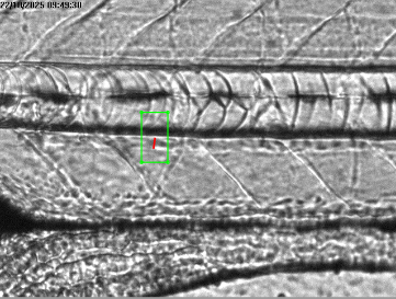
A Zebrafish Cardiovascular Analysis System is most useful when it turns a moving biological event into a reviewable research record. In a cardiac assay, a value such as heart rate is not enough on its own. Teams need to know which specimen was recorded, what the image showed, how the signal was extracted, and whether the conditions were comparable with prior runs. That is why modern platforms bring capture, measurement, and visualization into one workflow.
In zebrafish cardiovascular work, the practical challenge is often not collecting a video. It is connecting the video to a traceable review decision. A well-configured Zebrafish Cardiovascular Analysis System can support that connection by associating time-stamped imaging with calculated cardiac and vascular measures. The exact configuration should still fit the developmental stage, assay design, and imaging conditions in use.
Why real-time acquisition changes cardiovascular review
Cardiac function is dynamic. Contraction, relaxation, rhythm, and circulation change across successive frames, so the acquisition workflow needs that preserves the sequence rather than reducing it immediately to a single number. Time-lapse imaging has been used to record dynamic processes such as blood flow and heart contraction in zebrafish. In day-to-day work, that means the raw sequence remains available when a result needs to be checked.
Real-time acquisition also helps teams see whether the recording itself is suitable for review. A researcher can review focus, orientation, motion artifact, exposure, and the visibility of the cardiac region before accepting a measurement. This matters because an apparently neat result may reflect an unstable image sequence rather than a meaningful biological change. The workflow should therefore treat live review as a quality-control step, not merely as a display feature.
From image sequence to measurable parameters
The analytical value of an integrated platform comes from linking image data to clearly defined endpoints. Depending on the workflow, a laboratory may evaluate beat-to-beat timing, rhythm regularity, ventricular-wall motion, chamber dimensions, fractional shortening, vessel diameter, or visible blood-flow behavior. High-resolution imaging can also support hemodynamic measurements such as flow velocity in zebrafish embryos when the protocol and image quality are appropriate.
For a useful workflow, each metric needs a transparent calculation rule. Teams should document the region of interest, frame rate, measurement window, thresholding approach, and any exclusions. This is particularly important when data acquisition is automated: automation can reduce repetitive manual steps, but it does not remove the need to inspect edge cases. A trace that misses a weak contraction or includes body movement deserves review before it becomes a row in a final dataset.
Visualization is an review tool, not a presentation layer
Visualization lets the platform show how a reported value arose. Overlaying a tracked chamber boundary, motion curve, or flow path on the source image gives analysts a way to verify whether the software is following the intended anatomy. Trend plots can then make it easier to identify a gradual shift across a plate, a batch, or repeated acquisition sessions.
A useful lab habit is to inspect the video and overlay before opening the summary table. That takes a little longer up front, but it can avoid a much longer investigation later. The detail the camera catches can be different from what an observer notices during handling: a subtle orientation change, an out-of-plane heart, or a brief motion event may explain a surprising point. A platform that keeps visual evidence beside numerical outputs supports a more defensible decision process.
Build a comparable workflow around the system
The system performs within the boundaries set by the assay. Developmental stage, strain, temperature, anesthesia approach, illumination, magnification, and acclimation time can all affect what is visible and how results should be compared. Heart rate, for example, can adapt rapidly to temperature changes. The normal range is therefore not fixed in a way that makes a single threshold transferable between every experiment.
For that reason, a practical real-time workflow should record both the measurement and its context:
- Before capture: define stage, temperature target, plate layout, exposure conditions, and acceptance criteria.
- During acquisition: retain time stamps, specimen or well identity, frame rate, and a view suitable for assessing image quality.
- During analysis: apply the same regions of interest and calculation rules where the study design permits, while flagging exceptions rather than hiding them.
- During review: use visual overlays and time-series plots to investigate outliers, plate effects, and unexpected patterns.
- After review: export metrics with the linked acquisition metadata so another analyst can reconstruct the decision path.
This workflow helps the system contribute to reproducibility without claiming that one setup suits every study. If imaging-room temperature fluctuates or a developmental window changes, the comparison plan needs adjustment. Documenting those conditions is more informative than attempting to normalize every difference after the fact.
Where integrated acquisition and visualization help most
In early cardiovascular phenotyping, an integrated platform can help a team move from a visual observation to a structured set of measurements. In compound-screening workflows, it can make it easier to determine whether an apparent change is consistent across replicates and whether the source sequences support the reported pattern. In cardiotoxicity-oriented research, it can help reviewers examine rhythm, contractility, and flow-related observations together rather than treating them as isolated outputs.
There is an operational benefit as well. When several researchers contribute to a study, consistent capture settings and visible calculation rules make handoffs clearer. A system with traceable data acquisition can reduce ambiguity when one operator collects images and another reviews results. That does not replace training or protocol validation, but it gives both people the same evidence to inspect.
Questions to ask when evaluating a system
Rather than beginning with a feature checklist alone, evaluate how the system fits the full experimental loop. Ask whether it can preserve source imagery, identify specimens and acquisition conditions, display measurement overlays, export reviewable results, and support the endpoints your protocol actually requires. Also ask how the workflow handles low-quality recordings, missing metadata, or a result that conflicts with visual inspection.
For many teams, the useful shift is from collecting more numbers to creating evidence that can be interpreted in context. A system with real-time data acquisition and visualization can make that shift practical by keeping the image, the measurement, and the experimental context connected. When those elements remain visible together, researchers are better positioned to assess data quality, explain exceptions, and refine the next run.








