
 EN
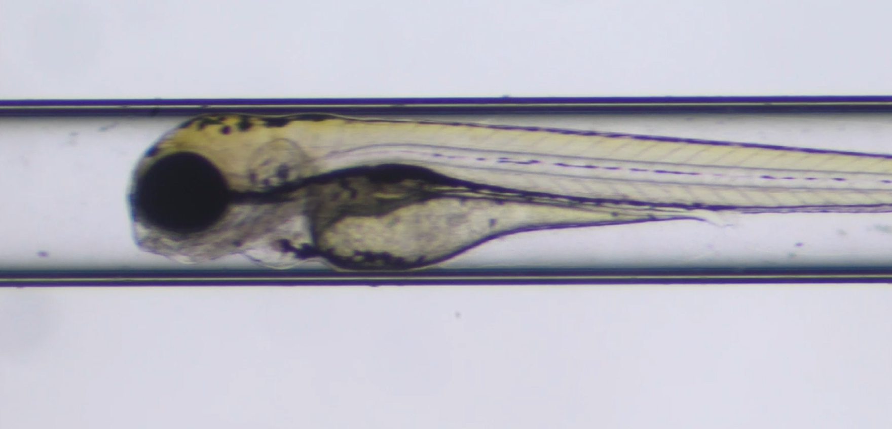
How Does a Zebrafish Imaging System Handle Multi-Well Plate Imaging?
A Zebrafish Imaging System turns a plate of living samples into a repeatable acquisition workflow. Rather than asking an operator to reposition every well and re-establish focus by hand, the system combines plate geometry, an automated stage, imaging recipes, and quality checks. That combination matters when a lab needs to compare embryos or larvae across treatments without letting small handling differences dominate the result.
In day-to-day zebrafish work, the hard part is rarely capturing one attractive image. The hard part is collecting comparable images from many wells while preserving orientation, exposure discipline, and sample traceability. A well-configured Zebrafish Imaging System makes that repeatability the center of the workflow.
What Multi-Well Plate Imaging Changes
A multi-well plate gives each sample a known physical address: plate format, row, column, and well. The imaging workflow uses those coordinates to create an acquisition map. The system can move to a selected well, identify or refine the imaging position, acquire the chosen channels, and record the result against that well address before moving on.
This is the foundation of high-content imaging. The aim is not simply more pictures; it is structured image data linked to treatment, time point, and readout. For a zebrafish assay, the readout might be morphology, fluorescent signal, movement, vascular features, or another phenotype defined in the study plan. The right measurement still depends on the biology and protocol. A faster scan does not automatically produce better data if larvae are poorly oriented or if focus varies from well to well.
How an Automated Stage Moves Through the Plate
The automated stage is the mechanical layer that translates a plate map into repeatable X-Y positions. After the plate is registered, the stage follows the stored well coordinates. A typical sequence is straightforward:
- Load the plate and confirm its format and orientation.
- Select the wells, fields, channels, and time points required by the assay.
- Move the automated stage to each planned well position.
- Use a focus routine or pre-set focal plane appropriate to the sample and objective.
- Capture images and save metadata with the plate and well identifiers.
- Flag wells that do not meet the study's quality criteria for review.
Motorized stage positioning is a recognized element of automated vertebrate screening workflows. In practice, its value is consistency: the same coordinate logic can be applied across a plate and repeated in later runs. Still, a stage does not correct upstream preparation problems. Bubbles, tilted plates, variable media depth, or animals settling at different depths can all change the focal plane and apparent phenotype.
Focus and Field Selection Are the Real Throughput Controls
For a Zebrafish Imaging System, well-to-well movement is only one part of throughput. The system also needs a plan for where to look inside each well. Depending on the plate, specimen size, and endpoint, that may mean one central field, several tiled fields, or a low-magnification overview followed by targeted higher-magnification captures.
Autofocus is useful when plate flatness or sample depth changes between wells, but it should be validated for the assay. For a long time it is tempting to treat the sharpest possible autofocus score as the goal; more consistent results often come from a focus rule that is robust to the relevant anatomy and fluorescent signal. If the imaging room, plate type, or anesthesia timing changes, re-check the focus strategy instead of assuming an old setting will transfer unchanged.
A sensible workflow also defines what counts as an acceptable field before acquisition begins. For example, a lab may require one embryo per well, an orientation range, a minimum signal level, or an unobstructed region of interest. Capturing those rules in the run setup reduces the temptation to make subjective field choices after results are visible.
Building a High-Content Acquisition Recipe
A repeatable high-content run is usually built from a recipe rather than from ad hoc controls. The recipe connects plate layout to acquisition settings and analysis expectations. It should state:
- Plate definition: format, well selection, and any wells reserved for controls.
- Optics: objective, illumination channel, exposure approach, and whether brightfield or fluorescence is needed.
- Stage plan: one field or multiple fields per well, plus any offset or tiling pattern.
- Focus method: fixed Z position, autofocus, or a validated hybrid approach.
- Timing: acquisition order and interval for time-lapse studies.
- Metadata: treatment identifiers, plate ID, well ID, time point, and run settings.
The details should remain proportionate to the assay. A development screen may prioritize coverage and timing, while a fluorescence assay may prioritize channel separation and exposure stability. There is no universal recipe; the right configuration depends heavily on specimen age, plate geometry, and the phenotype being measured.
Why Quality Control Must Happen During Imaging
Multi-well automation can create a large image set quickly, which makes early quality control more important, not less. A Zebrafish Imaging System should give operators a way to review representative wells and identify obvious problems such as empty wells, multiple animals, poor focus, saturated fluorescence, or unexpected debris.
Useful quality checks separate acquisition failures from biological results. If control wells show uneven illumination or if an entire row is out of focus, that is an imaging issue to investigate before interpreting treatment differences. Likewise, a clear naming and metadata convention makes it possible to trace an image back to its well, settings, and run. That traceability is especially valuable when a promising phenotype needs confirmation in a later plate.
Practical Setup Checklist for Multi-Well Zebrafish Imaging
- Confirm that the selected plate and holder are stable and correctly oriented.
- Use a plate map that makes controls and treatment groups easy to identify.
- Validate the automated stage coordinates on corner and center wells before a full run.
- Test focus and exposure on representative samples, including dim and bright conditions.
- Define what triggers a repeat image, an exclusion flag, or a manual review.
- Save the acquisition recipe with the raw images and analysis outputs.
Anyway, a useful first run is often a short pilot rather than a full plate. It reveals whether the imaging recipe matches the plate, specimen preparation, and endpoint. Once those pieces are aligned, the same Zebrafish Imaging System workflow can make multi-well plate imaging more comparable, more traceable, and easier to scale across routine studies.
Conclusion
A Zebrafish Imaging System handles multi-well plate imaging by linking a registered plate layout to automated movement, controlled focus, planned image acquisition, and well-level metadata. The automated stage provides the repeatable motion, while the high-content recipe and quality checks protect the meaning of the images. For laboratories planning a new assay, the key question is not only how many wells can be scanned, but whether each well is acquired under conditions that support a fair biological comparison.








