
 EN
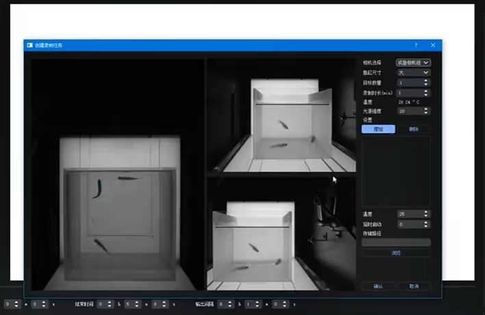
How does a zebrafish imaging system capture high-resolution images of live embryos?
A Zebrafish Imaging System produces useful images of live embryos by treating sample preparation, optics, illumination, timing, and analysis as one connected workflow. The camera matters, but it is only one part of the answer. An embryo that drifts, rotates, or experiences too much light can erase the practical value of an otherwise sharp frame.
From a zebrafish cardiovascular research perspective, image quality starts before acquisition. The goal is not simply to make a picture look crisp. It is to preserve a living embryo well enough that the resulting image can support a reproducible biological observation, whether the readout is morphology, fluorescent signal, cell movement, or a fast process such as cardiac motion.
What “high-resolution” means in live embryo imaging
In a Zebrafish Imaging System, high-resolution describes more than pixel count. Optical resolution determines how closely spaced details can be distinguished; sampling determines whether the detector records enough information to retain those details; contrast determines whether the feature can be separated from its background. For a live embryo, stability and viability belong in that same definition because motion blur, focal drift, and stress can hide structures that the optical train could otherwise resolve.
Zebrafish embryos are especially useful for imaging because their optical transparency permits direct observation during development. That advantage does not remove every imaging constraint. Pigmentation at later stages, tissue depth, fluorescence brightness, and the orientation of the region of interest all influence what is visible. A high-resolution result is therefore a fit between the biological question and the acquisition settings, not a single universal setting.
The capture chain inside a Zebrafish Imaging System
1. Prepare and orient the live embryo
The first practical task is controlled positioning. Live embryos may be mounted in a chosen orientation so the structure of interest faces the objective, and gentle immobilization helps reduce motion during exposure. Agarose-based mounting is widely used in long-term live-imaging workflows because it can hold orientation while leaving the embryo in an aqueous environment. The setup step nobody mentions enough is that a stable lateral view may be right for one phenotype, while a dorsal or ventral view may be needed for another.
A Zebrafish Imaging System benefits from a preparation that is repeatable across embryos. Consistent orientation makes focus, region-of-interest placement, and downstream comparison less ambiguous. It also prevents a common error: mistaking a changed viewing angle for a changed biological feature.
2. Deliver light without overwhelming the sample
Brightfield, transmitted light, fluorescence, confocal, spinning-disk, and light-sheet approaches create contrast in different ways. In every case, the illumination must be sufficient to capture the relevant signal while avoiding unnecessary exposure. More excitation light can raise signal in a short frame, but during a time series it may also increase photobleaching or phototoxicity. For live embryos, a technically sound Zebrafish Imaging System workflow usually begins with the lowest illumination and exposure that still answer the question.
This is where “high-resolution” needs a correction: a noisier but biologically gentle acquisition can be more valuable than a visually dramatic image that perturbs the embryo. Where that balance lands depends on fluorophore brightness, objective numerical aperture, camera sensitivity, frame rate, and the planned duration of the experiment.
3. Form a sharp image at the detector
Objectives collect light from the embryo and project it to a camera or scanning detector. Their numerical aperture, working distance, and correction for the mounting environment affect how much fine detail can be transferred into the image. Focus must be maintained at the target plane, especially when the embryo has depth or the experiment uses a z-stack. A Zebrafish Imaging System can capture a single plane for a surface-level question or collect multiple planes to reconstruct a thicker region.
Matching pixel size to optical resolution matters as well. If pixels are too large, fine detail is undersampled. If the acquisition samples far more finely than the optical system can resolve, data volume and light dose can increase without revealing proportionally more biology. In practice, the useful setting is the one that captures the relevant feature clearly enough for the planned measurement.
4. Control time and motion
Live embryos change while they are being imaged. A beating heart, circulation, locomotor movement, and developmental progression can each require different capture timing. A Zebrafish Imaging System may use short exposures, rapid frame sequences, triggered acquisition, or time-lapse intervals chosen around the biological event. For fast motion, an exposure that is too long can blur the structure even when focus is carefully maintained.
Anesthesia can reduce movement in some protocols, but it is not a fixed target that can be copied blindly between stages or assays. Dose, exposure time, temperature, developmental stage, and the endpoint all matter. The image actually shows the embryo under those conditions, so documenting them is part of interpreting the result.
How high-resolution capture becomes reliable data
A Zebrafish Imaging System produces measurement-ready data when acquisition choices are paired with consistent metadata and analysis rules. Before comparing groups, a lab can define the orientation, magnification, illumination channel, exposure, z-step, time interval, and analysis region. That consistency helps distinguish a real phenotype from an acquisition difference.
- For morphology: keep scale, orientation, and focus criteria consistent before measuring length, area, or shape.
- For fluorescence: avoid changing exposure or gain between groups unless the analysis plan accounts for it.
- For dynamic events: select a frame rate that can capture the event without excessive motion blur or light dose.
- For 3D data: set z-spacing and depth coverage according to the structure, rather than collecting a larger stack by default.
Automation can make a Zebrafish Imaging System more consistent by standardizing loading, positioning, focus routines, and image naming. It does not remove the need for validation. Automated segmentation or phenotype calls should be checked against representative raw images, particularly when developmental stage, orientation, or signal quality varies.
A practical checklist before you capture
- State the biological feature and the smallest detail that must be distinguished.
- Choose an embryo orientation that exposes that feature to the optical path.
- Confirm stable mounting and a viable aqueous environment.
- Set illumination and exposure conservatively, then verify that the signal is adequate.
- Test focus, frame rate, and z-coverage on a small pilot set before scaling the run.
- Record settings so another session can reproduce the capture conditions.
The bottom line
A Zebrafish Imaging System captures high-resolution images of live embryos by coordinating biological preparation with optical acquisition and analysis. Transparency creates an opportunity to observe development directly, but the usable detail comes from disciplined control of orientation, focus, illumination, timing, and metadata. When those parts are aligned, a high-resolution image becomes more than an attractive frame: it becomes evidence that can be compared, quantified, and interpreted with appropriate care.








