
 EN
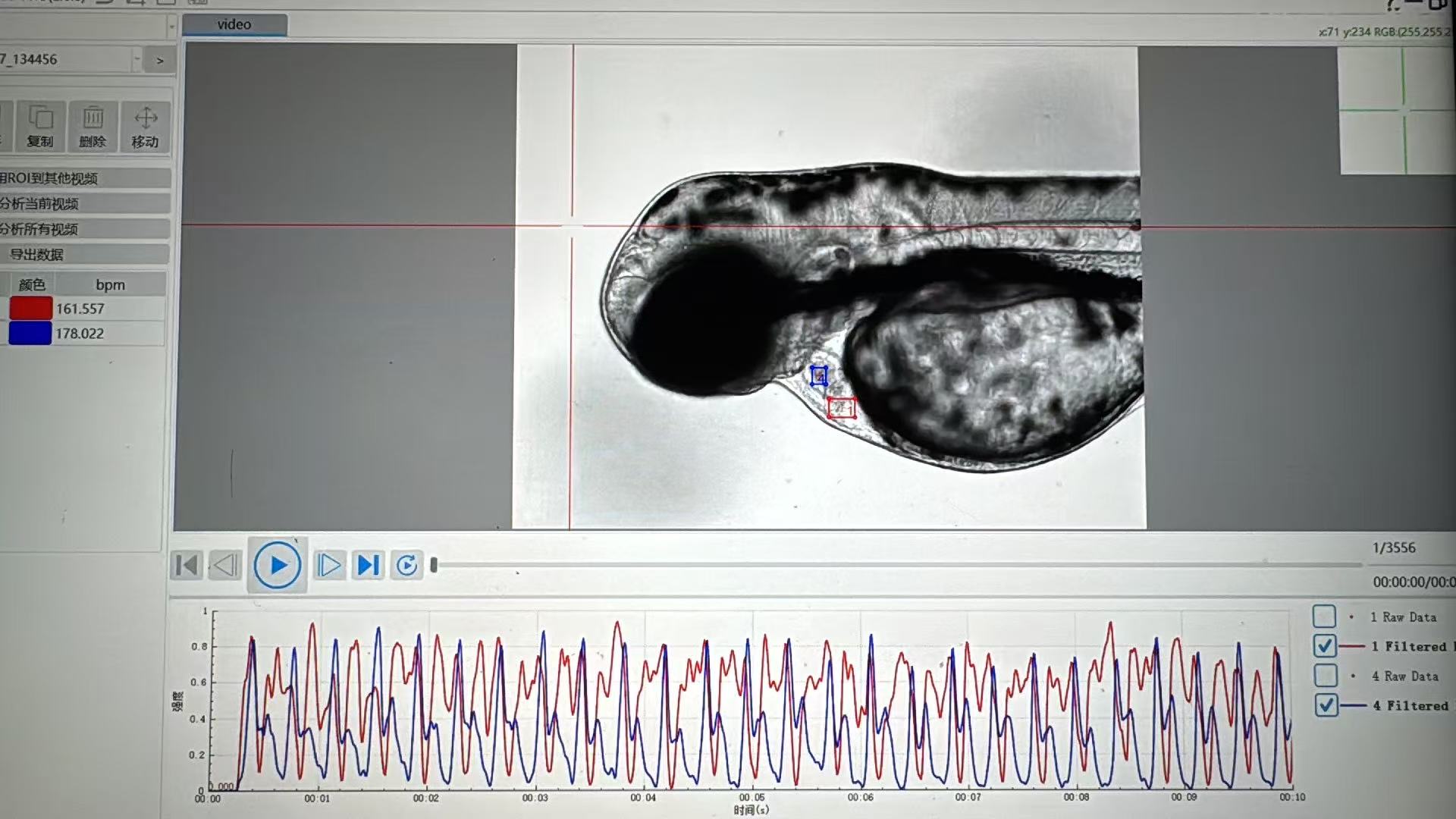
How Does a Zebrafish Cardiovascular Analysis System Compensate for Motion During Imaging?
A zebrafish heart is a moving target by design. In a short video, the ventricle contracts, the atrium follows, blood cells change position, and the entire larva can shift slightly in the field of view. A useful Zebrafish Cardiovascular Analysis System therefore does more than record a movie. It separates biologically meaningful motion from unwanted image displacement so that heart rate, rhythm, chamber motion, and flow-related readouts are measured from comparable frames.
As researchers who work with zebrafish cardiovascular studies know, the important question is not whether motion exists; it is expected. The practical question is whether the workflow can identify the cardiac cycle, maintain the region of interest, and flag footage that is too unstable for a trustworthy result. Motion compensation is a chain of acquisition and analysis decisions, not a cosmetic filter applied at the end.
Why motion creates an analysis problem
In zebrafish imaging, several movements may occur at once. The heartbeat is the signal of interest, while whole-body translation, rotation, focus drift, vibration, and changes in illumination can be noise. If a chamber boundary appears to move because the larva slid by a few pixels, an algorithm may mistake that shift for contraction. If the region of interest drifts, a fixed measurement window can clip the heart or include surrounding tissue. Either error can reduce accuracy even when the recorded video looks acceptable to the eye.
This distinction matters especially in repeated imaging. A small alignment error can be tolerable for a single qualitative observation but become important when a lab compares treatment groups, time points, or many larvae. The most dependable workflows control motion before acquisition and then use software to correct the residual movement that remains.
Layer 1: reduce motion before the camera starts
Good stabilization begins with sample preparation. The larva should be positioned consistently, the imaging chamber should minimize unintended drift, and illumination and focus should be stable before the recording begins. Researchers also need a repeatable policy for developmental stage, temperature, acclimation, and exposure settings, because those conditions can change cardiac behavior as well as image quality.
This is a useful scope limit: software cannot reliably recover a video in which the heart leaves the field, is persistently blurred, or is obscured by a poor orientation. A Zebrafish Cardiovascular Analysis System can improve alignment of usable frames, but it should not turn low-quality acquisition into an apparently precise measurement. In practice, documenting a rejection rule is as important as choosing a correction algorithm.
Layer 2: register frames to a stable reference
After capture, the system can estimate non-cardiac displacement between frames. A reference image or reference region is selected, often using stable features near the heart rather than the contracting chamber alone. The analysis then calculates how later frames must be translated—and, where the workflow supports it, rotated or otherwise aligned—to match that reference. The corrected sequence keeps the field of view steady while preserving the local deformation caused by the heartbeat.
For example, if the entire larva moves slightly to the right, frame registration shifts the image back before chamber measurements are made. That operation is different from smoothing the heart itself. The goal is not to erase contraction; it is to prevent global movement from being counted as contraction. A careful workflow keeps the original video available for review and records whether correction was applied.
Choosing the reference region matters
Registration is more reliable when its reference contains features that remain recognizable from frame to frame. Using a broad, high-contrast area around the heart can be more robust than anchoring on a boundary that changes dramatically during systole. If the reference includes too much moving anatomy, the correction may follow the heartbeat instead of the larva. This is one reason a visual overlay or before-and-after playback is valuable during method setup.
Layer 3: align frames by cardiac phase
Image registration corrects specimen drift, but it does not make different points in the heartbeat equivalent. Cardiac phase alignment addresses that separate issue. A system can derive a repeating signal from chamber appearance, brightness changes, motion traces, or a selected region of interest, then assign each frame to a phase of the cardiac cycle. Frames captured at similar phases can be grouped for comparison or reconstruction.
Retrospective optical gating is one example of this principle: a sequence spanning multiple beats is processed so that frames are labeled and sorted by phase. In longer experiments, adaptive approaches may update the reference as the developing heart changes. For routine video analysis, the same logic helps a Zebrafish Cardiovascular Analysis System compare like with like—end-diastolic frames with end-diastolic frames, and systolic frames with systolic frames—instead of averaging incompatible moments together.
Layer 4: track the heart rather than a fixed box
Once global motion is reduced, the analysis can maintain a heart-centered region of interest. Tracking may use contrast, boundary shape, template similarity, or segmentation to relocate the chamber area from frame to frame. The tracked region allows measurements to follow the heart when there is modest residual drift. It also helps keep atrial and ventricular signals distinct when the system calculates cycle timing or relative chamber motion.
Tracking is not a substitute for sensible quality control. If the predicted region jumps unpredictably, overlaps another structure, or loses the target, the system should flag the sequence for review rather than silently filling gaps. The metric that changes a protocol is often not the mean heart rate; it is the proportion of recordings that pass a clear tracking and focus check.
How compensation protects measurement accuracy
Motion compensation supports accuracy by making each measurement refer to the same anatomy and the same phase of motion. For heart rate, that means peak detection is less likely to respond to camera shake. For rhythm assessment, it makes cycle-to-cycle timing less vulnerable to abrupt ROI shifts. For contractility-oriented measures, it reduces the chance that apparent displacement of a chamber wall is caused by whole-animal translation. For flow-oriented imaging, stable alignment makes it easier to evaluate changing blood-cell patterns within a consistent vessel or cardiac region.
Still, correction should be conservative. Excessive smoothing, poorly chosen registration features, or aggressive interpolation can alter edges and timing. The right approach depends on frame rate, magnification, sample orientation, and the metric being reported. In other words, motion compensation should be validated against the specific assay rather than treated as a universal setting.
A practical workflow for reliable imaging
- Standardize acquisition. Set specimen orientation, focus, exposure, and environmental conditions before recording.
- Record enough temporal detail. Use a frame rate appropriate for resolving the cardiac motion needed by the planned metric.
- Inspect raw footage. Exclude clips with sustained blur, major specimen displacement, or an obscured heart.
- Apply global registration. Align residual field movement to stable image features while preserving cardiac deformation.
- Track the cardiac region. Confirm that the region of interest stays on the intended anatomy across the sequence.
- Assign or verify cardiac phase. Compare frames from equivalent points in the cycle when reconstructing or measuring motion.
- Review correction outputs. Check overlays, tracking confidence, and flagged clips before accepting the final values.
What to ask when evaluating a system
When evaluating a Zebrafish Cardiovascular Analysis System, ask whether its workflow distinguishes global specimen movement from heart-wall motion, how it maintains or updates regions of interest, and what evidence it provides when tracking fails. It is also sensible to ask how raw and corrected videos are retained, whether correction settings are documented, and whether the analysis can be reviewed at the frame level. These questions make it easier to defend a result during method development, internal review, or later comparison across runs.
Hunter Lab focuses on zebrafish instruments and applications, so laboratories can discuss the imaging conditions and cardiovascular endpoints that matter for their own studies. No single compensation method fits every assay. A suitable choice preserves the biology, exposes uncertainty, and produces a repeatable path from raw imaging to interpretable data.
Conclusion
A Zebrafish Cardiovascular Analysis System compensates for motion by combining physical stabilization with frame registration, cardiac-phase alignment, heart-region tracking, and explicit quality checks. Together, these steps keep unintended drift from being interpreted as physiology while preserving the contraction and flow changes that the experiment is meant to measure. For dependable imaging, validate the full workflow on representative recordings and treat failed tracking or poor acquisition as information to review, not data to hide.








