
 EN
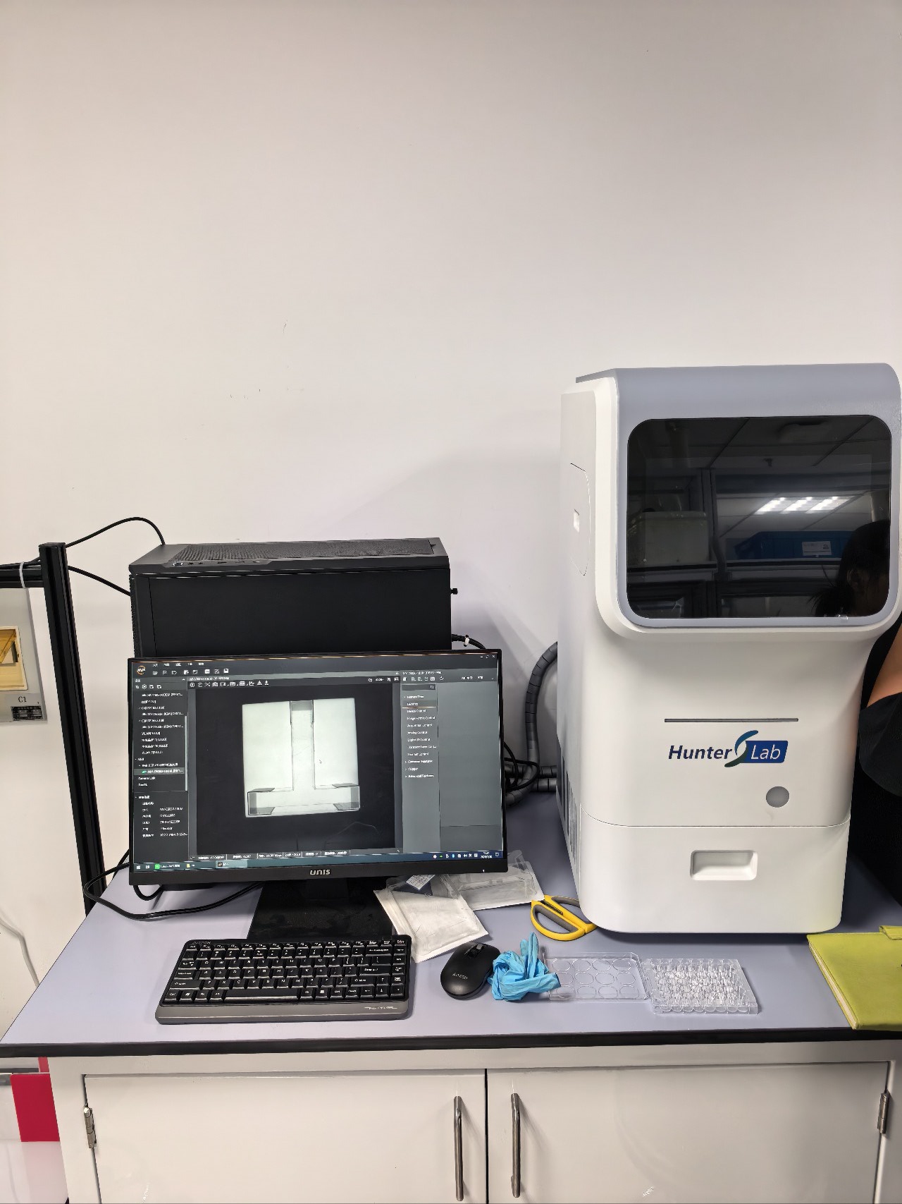
A zebrafish imaging system enables non-invasive observation of internal organ development.
For developmental biologists, the appeal of a Zebrafish Imaging System is straightforward: it can turn changes that would otherwise be inferred from an endpoint into observations made in a living organism. Early zebrafish embryos are optically accessible, so researchers can follow the formation and behavior of selected internal organs without opening the animal or collecting tissue at every timepoint. That does not make imaging effortless. It makes the experimental question, the developmental stage, and the acquisition plan more important.
A useful Zebrafish Imaging System is therefore more than a camera. It is a coordinated workflow for holding a sample, selecting contrast, capturing a repeatable view, and preserving the context needed to interpret change over time. For a graduate researcher planning a first longitudinal study, that distinction often prevents a technically attractive image from becoming biologically ambiguous data.
Why zebrafish support non-invasive developmental observation
During early life, zebrafish embryos and larvae offer a practical optical window into development. Brightfield imaging can show gross morphology, while fluorescent reporters, stains, or other contrast methods can help isolate particular cells, tissues, or processes. A Zebrafish Imaging System can support either approach when its optics, illumination, and acquisition settings match the question being asked.
The key word is non-invasive, not consequence-free. A non-invasive image does not automatically eliminate handling stress, motion, phototoxicity, anesthetic effects, or temperature drift. The advantage is that the same cohort may remain available for follow-up. Instead of comparing one group at an early stage with a different group later, researchers can often observe trajectories within an appropriately managed experimental design.
This is especially helpful when the question concerns the sequence rather than the final appearance of internal organs. A Zebrafish Imaging System can document when a structure first becomes distinguishable, how its shape changes, or whether a reporter pattern expands, contracts, or shifts location. The image is evidence of what was visible under defined conditions; it is not, by itself, proof of mechanism.
Start with the organ question, not the instrument menu
Before selecting settings on a Zebrafish Imaging System, write the decision that each image should help make. Is the study asking whether cardiac morphology changes after exposure? Whether renal development proceeds on schedule? Whether a fluorescent signal is localized to a defined region? Each question has a different tolerance for field of view, depth, resolution, time interval, and sample throughput.
- Whole-animal context: Use broad views when relative position and overall morphology matter.
- Organ-focused morphology: Use a stable orientation and sufficient resolution when the boundary or shape of an organ is the readout.
- Dynamic function: Use acquisition timing that can resolve the event of interest, then document the frame rate and analysis rule.
- Reporter-based observation: Keep illumination and exposure comparable so apparent signal changes are not only acquisition changes.
A Zebrafish Imaging System should be assessed against these requirements, not against the longest specification list. More magnification does not necessarily produce more useful data; it may reduce field of view, increase sensitivity to movement, and slow the workflow. Similarly, an automated workflow can reduce repetitive handling, but it still needs a plan for orientation, focus failures, and samples that fall outside the expected pattern.
Build a repeatable acquisition workflow
In practice, the most valuable feature of a Zebrafish Imaging System is repeatability. Decide in advance how larvae will be staged, transferred, oriented, and identified. Record the optical mode, objective, illumination, exposure, acquisition interval, temperature conditions, and any immobilization or anesthesia procedure. These details may feel procedural, but they explain whether two image series are comparable.
Stage selection deserves particular attention. Organ formation and visibility can change rapidly during early development, so a nominal age alone may not be enough when embryos progress at different rates. Define the staging criteria that matter to the study and apply them consistently. If imaging spans several days, use the same landmarks and naming conventions at every session. A Zebrafish Imaging System can make repeated observation practical, but it cannot correct a timeline that was not defined.
It also helps to include controls that reveal acquisition drift. Reference samples, consistent plate positions where feasible, and a small review set of manually checked images can show whether an apparent change comes from biology or the workflow. This matters for internal organs whose contrast may be subtle. What looks like a new phenotype can sometimes be a focus shift, a changed viewing angle, or a difference in the degree of immobilization.
Use images to connect morphology with time
The value of a Zebrafish Imaging System grows when images are organized as a time-resolved record rather than a gallery. A simple schedule might capture a baseline image, one or more defined observation windows, and a final follow-up. The interval should be driven by the rate of the biological process, not by the convenience of the workstation.
For example, a study of organ development may pair whole-body images with a closer organ view at each timepoint. The first series preserves context; the second provides a consistent location for measurements or blinded scoring. The approach is non-invasive in the sense that it can preserve the cohort for later observation, provided the protocol has been tested for the relevant sample and readout.
Analysis should be planned at the same time as imaging. Define what will be scored, measured, or classified before looking at the full dataset. A Zebrafish Imaging System may generate many images quickly, but quantity does not replace a clear endpoint. When automated analysis is used, review representative outputs across control and test conditions, especially near the boundaries where segmentation or classification could fail.
Questions to ask when evaluating a Zebrafish Imaging System
- Can the Zebrafish Imaging System produce the optical contrast needed for the target organ and stage?
- Can it maintain a repeatable sample orientation and focus for the required observation window?
- Does its acquisition speed match the process being observed without creating unnecessary light exposure?
- Can its data files, metadata, and image-analysis outputs be linked to individual samples and timepoints?
- Can the workflow be validated with known normal and altered phenotypes before a large experiment begins?
The answers will vary across projects. A system optimized for high-throughput morphology may not be a suitable choice for a deep, high-resolution study, and a strong single-sample setup may not scale cleanly to screening. That is not a flaw; it is a reason to make the biological question explicit.
A measured path from image to interpretation
A Zebrafish Imaging System enables a powerful kind of observation: watching living internal organs change across defined stages instead of relying solely on terminal snapshots. Its strongest contribution is not simply visual appeal. It is the ability to collect comparable, staged evidence while keeping interpretation tied to controls, handling conditions, and a pre-specified endpoint.
For researchers working on developmental questions, begin with a small pilot. Test visibility, sample stability, image quality, and recovery after imaging. Then scale the workflow only after the images answer the intended question reliably. Used this way, a Zebrafish Imaging System supports careful, non-invasive observation without promising more than the experiment can demonstrate.








